This site uses cookies to improve your experience. To help us insure we adhere to various privacy regulations, please select your country/region of residence. If you do not select a country, we will assume you are from the United States. Select your Cookie Settings or view our Privacy Policy and Terms of Use.
Cookie Settings
Cookies and similar technologies are used on this website for proper function of the website, for tracking performance analytics and for marketing purposes. We and some of our third-party providers may use cookie data for various purposes. Please review the cookie settings below and choose your preference.
Used for the proper function of the website
Used for monitoring website traffic and interactions
Cookie Settings
Cookies and similar technologies are used on this website for proper function of the website, for tracking performance analytics and for marketing purposes. We and some of our third-party providers may use cookie data for various purposes. Please review the cookie settings below and choose your preference.
Strictly Necessary: Used for the proper function of the website
Performance/Analytics: Used for monitoring website traffic and interactions
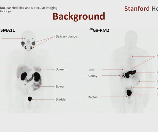
such as Ga-68 PSMA-11 and F-18 DCFPyl (Pylarify, Lantheus MedicalImaging) and are included in National Comprehensive Cancer Network guidelines, Duan explained. ng/mL after prostatectomy or ≥ 2 ng/mL after radiotherapy) and negative conventional imaging (CT and/or bonescans).
While broken bones are common injuries experienced across all ages, older individuals are at higher risk. Bones become more fragile and porous with age, so less force is needed to break their structure. Medicalimaging is used to help diagnose these injuries, so doctors can propose appropriate treatment plans.
To design the salvage radiation therapy, imaging is required to detect and locate the recurrence disease regime.” This practice, while meticulous, is labor-intensive and prone to inter- and intra-observer variations.
This scan may be performed in order to check whether you have a suspected kidney obstruction or impaired function. 3-Phase BoneScan A 3-phase bonescan is used to diagnose abnormal areas within the bones or joints, such as a fracture, bone pain, infection, or bone disease, which cannot be seen successfully with a standard X-ray.
MRI technologies are also particularly beneficial for scanning the brain, spine, soft tissues in the joints, and the interior structures of bones. The post Magnetic Resonance Imaging (MRI): A Leading Imaging Modality Because of its Diagnostic Versatility appeared first on Associates in MedicalImaging.
Capitol Imaging Services offers a variety of ultrasound imaging tests, like: Liver Elastography Ultrasound Guided Breast Biopsy Nuclear Medicine Nuclear Medicine imagingscans utilize a small, harmless dose of a radioactive isotope to highlight parts of the body, making them easier to see in an imaging test.
A bone density test is a non-invasive procedure to measure the strength of your bones. It is an effective way to diagnose bone-related health problems, such as osteoporosis, and to assess the risk of developing them.
We organize all of the trending information in your field so you don't have to. Join 5,000 users and stay up to date on the latest articles your peers are reading.
You know about us, now we want to get to know you!
Let's personalize your content
Let's get even more personalized
We recognize your account from another site in our network, please click 'Send Email' below to continue with verifying your account and setting a password.


Let's personalize your content