This site uses cookies to improve your experience. To help us insure we adhere to various privacy regulations, please select your country/region of residence. If you do not select a country, we will assume you are from the United States. Select your Cookie Settings or view our Privacy Policy and Terms of Use.
Cookie Settings
Cookies and similar technologies are used on this website for proper function of the website, for tracking performance analytics and for marketing purposes. We and some of our third-party providers may use cookie data for various purposes. Please review the cookie settings below and choose your preference.
Used for the proper function of the website
Used for monitoring website traffic and interactions
Cookie Settings
Cookies and similar technologies are used on this website for proper function of the website, for tracking performance analytics and for marketing purposes. We and some of our third-party providers may use cookie data for various purposes. Please review the cookie settings below and choose your preference.
Strictly Necessary: Used for the proper function of the website
Performance/Analytics: Used for monitoring website traffic and interactions
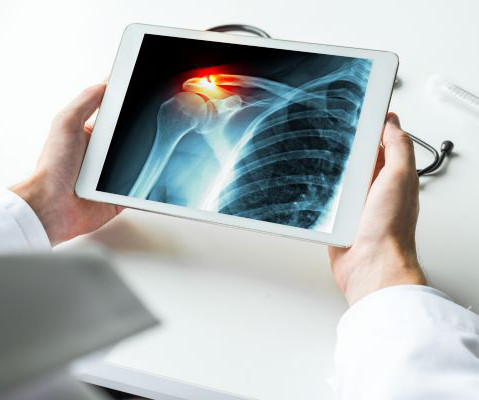
While broken bones are common injuries experienced across all ages, older individuals are at higher risk. Bones become more fragile and porous with age, so less force is needed to break their structure. Medicalimaging is used to help diagnose these injuries, so doctors can propose appropriate treatment plans.
Nuclear medicine tests use a very small amount of radioactive tracer (radionuclide or radioisotope) which is specific for the organ or tissue to be scanned. The gamma rays emitted by the tracer are detected by a special camera that is positioned near the organ or tissues being imaged.
There are several types of imaging tests that physicians use to detect cancer in patients: X-Ray, Computed Tomography (CT), Magnetic Resonance Imaging (MRI), Ultrasound (US), Nuclear Medicine, and Positron Emission Tomography (PET). It is also used to determine the progression of existing cancerous masses.
There are no special instructions to prepare for it, and unlike an MRI or CT Scan , a bone densitometry procedure does not involve lying in an enclosed tunnel. Instead, you will lie still on an X-ray table as the scanner passes over your body.
MRI technologies are also particularly beneficial for scanning the brain, spine, soft tissues in the joints, and the interior structures of bones. The post Magnetic Resonance Imaging (MRI): A Leading Imaging Modality Because of its Diagnostic Versatility appeared first on Associates in MedicalImaging.
We organize all of the trending information in your field so you don't have to. Join 5,000 users and stay up to date on the latest articles your peers are reading.
You know about us, now we want to get to know you!
Let's personalize your content
Let's get even more personalized
We recognize your account from another site in our network, please click 'Send Email' below to continue with verifying your account and setting a password.


Let's personalize your content