This site uses cookies to improve your experience. To help us insure we adhere to various privacy regulations, please select your country/region of residence. If you do not select a country, we will assume you are from the United States. Select your Cookie Settings or view our Privacy Policy and Terms of Use.
Cookie Settings
Cookies and similar technologies are used on this website for proper function of the website, for tracking performance analytics and for marketing purposes. We and some of our third-party providers may use cookie data for various purposes. Please review the cookie settings below and choose your preference.
Used for the proper function of the website
Used for monitoring website traffic and interactions
Cookie Settings
Cookies and similar technologies are used on this website for proper function of the website, for tracking performance analytics and for marketing purposes. We and some of our third-party providers may use cookie data for various purposes. Please review the cookie settings below and choose your preference.
Strictly Necessary: Used for the proper function of the website
Performance/Analytics: Used for monitoring website traffic and interactions
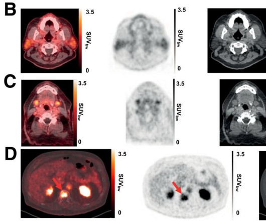
FDG-PET/CT provides functional insights by detecting metabolic changes before structural alterations, making it valuable for therapy response monitoring, but it has limitations in visualizing metabolically silent bone metastases, according to Masperi and Girlando.
In the trial, 30 patients with recurrent disease or metastatic prostate cancer underwent baseline conventional imaging (CT, MRI, or bonescan) and whole-body Ga-68 PSMA-R2 PET/CT between 20 minutes and four hours after injection. All patients had undergone either radical prostatectomy or radiation therapy. (A)
Imagine going in for a diagnostic MRIscan and coming out with a progressive, irreversible, and potentially fatal disease. Last week, attorneys for a Cleveland man announced they had filed suit against a gadolinium manufacturer, claiming that he developed NSF from an injection of gadolinium MRI contrast. 356: 9234, pp.
mtaschetta-millane Tue, 07/02/2024 - 09:53 July 2, 2024 — A new editorial paper was published in Oncoscience ( Volume 11 ) on May 20, 2024, entitled, “ Deep learning-assisted lesion segmentation in PET/CT imaging: A feasibility study for salvage radiation therapy in prostate cancer.” In this new editorial, researchers Richard L.J.
Bones become more fragile and porous with age, so less force is needed to break their structure. While X-rays are typically utilized, an MRI or CT scan may be recommended. What Are Broken Bones? To determine if the bone is broken, doctors may use one of the following imaging technologies.
Although the patient is exposed to radiation during the procedure, the test is considered safe because the amount of radiation used is comparable to that of a routine X-ray. This scan may be performed in order to check whether you have a suspected kidney obstruction or impaired function.
There are no special instructions to prepare for it, and unlike an MRI or CT Scan , a bone densitometry procedure does not involve lying in an enclosed tunnel. The scan uses a very low dose of radiation to produce pictures of the inside of the body, particularly around the hips and lower spine, to measure bone mass and strength.
We organize all of the trending information in your field so you don't have to. Join 5,000 users and stay up to date on the latest articles your peers are reading.
You know about us, now we want to get to know you!
Let's personalize your content
Let's get even more personalized
We recognize your account from another site in our network, please click 'Send Email' below to continue with verifying your account and setting a password.


Let's personalize your content