This site uses cookies to improve your experience. To help us insure we adhere to various privacy regulations, please select your country/region of residence. If you do not select a country, we will assume you are from the United States. Select your Cookie Settings or view our Privacy Policy and Terms of Use.
Cookie Settings
Cookies and similar technologies are used on this website for proper function of the website, for tracking performance analytics and for marketing purposes. We and some of our third-party providers may use cookie data for various purposes. Please review the cookie settings below and choose your preference.
Used for the proper function of the website
Used for monitoring website traffic and interactions
Cookie Settings
Cookies and similar technologies are used on this website for proper function of the website, for tracking performance analytics and for marketing purposes. We and some of our third-party providers may use cookie data for various purposes. Please review the cookie settings below and choose your preference.
Strictly Necessary: Used for the proper function of the website
Performance/Analytics: Used for monitoring website traffic and interactions
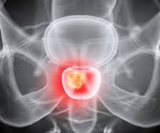
To address this gap in evidence, the researchers developed a decision analytic model to estimate clinical outcomes associated with PSMA-PET versus conventional imaging strategies, namely CT and bonescans.
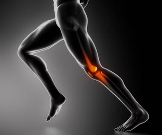
In an analysis of x-rays and bonescans, a group from Erasmus Medical Center in Rotterdam, the Netherlands, found that weight-bearing recreational activity was significantly associated with increased odds of knee osteoarthritis in individuals with low levels of lower-limb muscle mass.
At Clermont Radiology’s Women’s Center, we offer Digital Mammography, Ultrasound, and DEXA Scans. Below is a description of each type of exam: Digital Mammography Mammography is the practice of radiographic imaging of the soft tissue of the breast. Digital Mammography is the highest image resolution available.
Findings include: enhancement (hypointense on T1 and hyperintense on T2) of vertebral endplates and adjacent disc space (Image 1) CT Scan with IV contrast – use only of MRI contraindicated Inferior in evaluation of disc spaces and neural tissues Less sensitive than MRI and may be falsely negative in early disease Used primarily by surgeons for biopsy (..)
We organize all of the trending information in your field so you don't have to. Join 5,000 users and stay up to date on the latest articles your peers are reading.
You know about us, now we want to get to know you!
Let's personalize your content
Let's get even more personalized
We recognize your account from another site in our network, please click 'Send Email' below to continue with verifying your account and setting a password.


Let's personalize your content