Remove Clinic Remove Computed Tomography Remove Global Radiology
article thumbnail

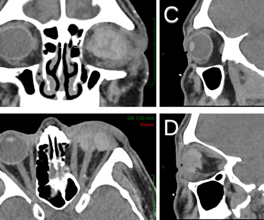
Ruptured Globe

Global Radiology CME

1 Imaging: Computed tomography (CT) is the recommended imaging modality for evaluating orbital trauma. Clinical features of single and repeated globe rupture after penetrating keratoplasty. Accuracy of Computed Tomography Imaging Criteria in the Diagnosis of Adult Open Globe Injuries by Neuroradiology and Ophthalmology.

article thumbnail

Acute Infective Vasitis

Global Radiology CME

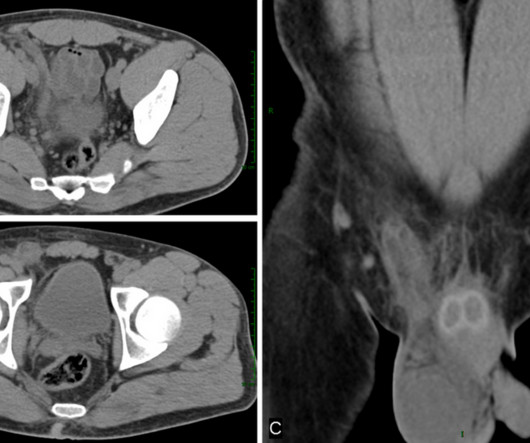
Vasitis: a clinical confusion diagnosis with inguinal hernia. Vasitis: clinical and ultrasound confusion with inguinal hernia clarified by computed tomography. Clinical Radiology. Vasitis: the need for imaging and clinical acumen. References: Yang DM, Kim HC, Lee HL, Lim JW, Kim GY. Int Braz J Urol.

Insiders

Sign Up for our Newsletter

This site is protected by reCAPTCHA and the Google Privacy Policy and Terms of Service apply.

Trending Sources

article thumbnail

Lisfranc Fracture Dislocation

Global Radiology CME

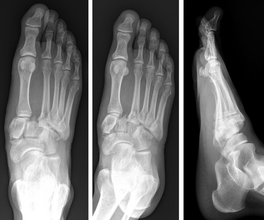
A computed tomography scan will better assist with diagnosis and help with planning if surgery is necessary [3]. Utility of weight-bearing radiographs compared to computed tomography scan for the diagnosis of subtle Lisfranc injuries in the emergency setting. J Foot Ankle Res. 2023;16(1):9. Published 2023 Mar 1.

article thumbnail

Capsule Retention Following Capsule Endoscopy

Global Radiology CME

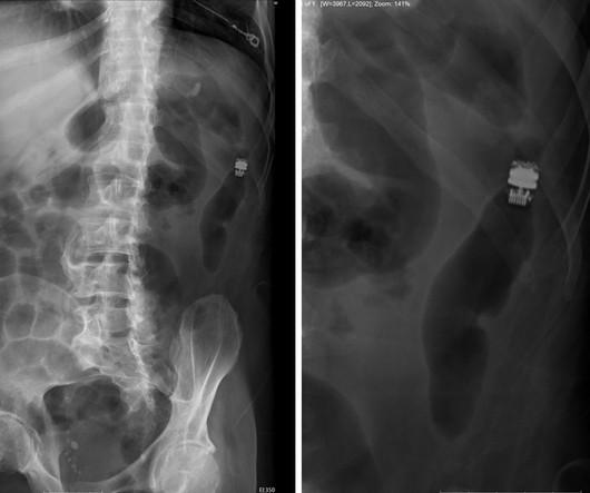
Approximately 2% of all capsule endoscopies result in CR [ 2 ] The clinical indication for capsule endoscopy is correlated with different rates of CR. Computed tomography (CT) could be used to determine the capsule’s location if it is difficult to do so via x-ray. Fernández-Urién I, Carretero C, González B, et al.

X-ray 52
article thumbnail

Lens Subluxation

Global Radiology CME

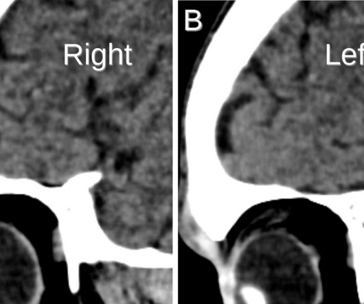
Computed tomography is also an alternative method for lens subluxation which again can show deviation of the lens (Figs. Sai is also a member of the Gold Humanism Honor Society and is involved with giving back to the community at a local free clinic as a medical assistant.

article thumbnail

Emphysematous Pyelonephritis

Global Radiology CME

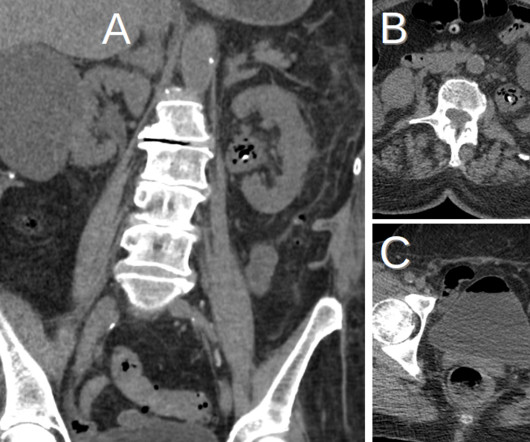
KUB indicates kidneys, ureter, and bladder (plain abdominal radiograph); CT, computed tomography; and PCD, percutaneous catheter drainage. In severe cases or patients who do not respond to PCD, treatment with nephrectomy can lead to clinical and radiological improvement (Fig. doi: 10.1148/radiology.198.2.8596845