This site uses cookies to improve your experience. To help us insure we adhere to various privacy regulations, please select your country/region of residence. If you do not select a country, we will assume you are from the United States. Select your Cookie Settings or view our Privacy Policy and Terms of Use.
Cookie Settings
Cookies and similar technologies are used on this website for proper function of the website, for tracking performance analytics and for marketing purposes. We and some of our third-party providers may use cookie data for various purposes. Please review the cookie settings below and choose your preference.
Used for the proper function of the website
Used for monitoring website traffic and interactions
Cookie Settings
Cookies and similar technologies are used on this website for proper function of the website, for tracking performance analytics and for marketing purposes. We and some of our third-party providers may use cookie data for various purposes. Please review the cookie settings below and choose your preference.
Strictly Necessary: Used for the proper function of the website
Performance/Analytics: Used for monitoring website traffic and interactions
Middlebrooks, MD, Mayo Clinic, Jacksonville, FL Erik H. Middlebrooks, MD, of the Mayo Clinic in Jacksonville, FL. Given the extensive array of sequences and techniques employed in clinical imaging, we must address the inherent challenges associated with each," he noted. I'm a radiographer,' " Stewart recalled.
Together with our radiographers, I learned to scan cardiac patients and learned special anatomy from pediatric cardiologists and pediatric cardiac surgeons." International organizations such as the Society of Cardiac ComputedTomography (SCCT) and the Society for Cardiac Magnetic Resonance (SCMR) also offer certifications.
S1-SSCH01-5 | E451A This scientific paper may increase overall confidence in the potential of using multimodal AI for tuberculosis (TB) detection, and potentially autonomous reporting, on chest radiographs in certain clinical settings. First PANORAMA findings support AI-based opportunistic screening Sunday, December 1 | 10:40 a.m.-10:50
Most Influential Radiology Researcher Erik Middlebrooks, MD, Mayo Clinic, Jacksonville, FL Erik Middlebrooks, MD. One of this year's nominations for Most Influential Radiology Researcher is Erik Middlebrooks, MD, of the Mayo Clinic in Jacksonville, FL.
Detecting CSIs in a clinical setting often requires imaging such as X-rays and computedtomography (CT) scans, both of which expose children to radiation, which can cause other health issues over time.
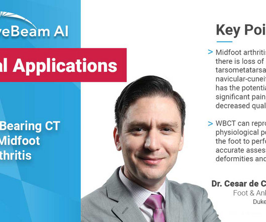
Diagnosis An accurate diagnosis and grading of the severity of osteoarthritic joints in the midfoot have been shown to be clinically relevant in treating the pathology early in its course and avoiding late-stage invasive procedures such as arthrodesis 3. Images demonstrated severe degeneration of the midfoot joints. PMID: 33504217. (4)
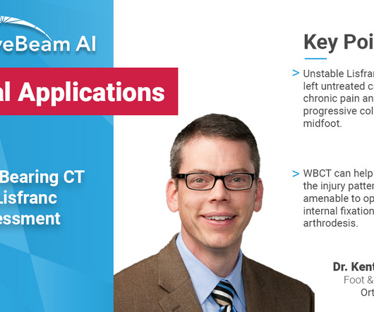
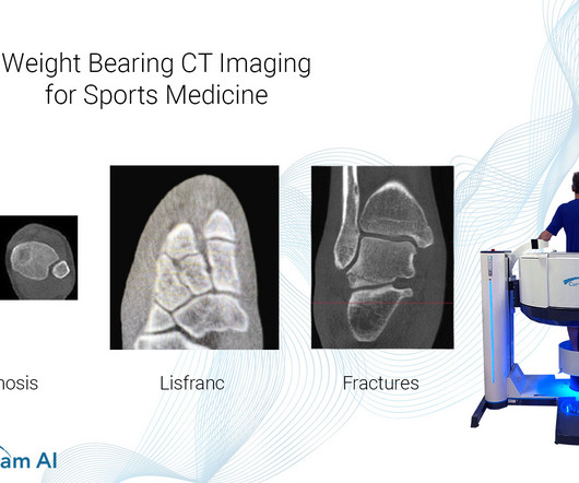
Diagnosis The diagnosis of Lisfranc injuries may be challenging on plain radiographs alone. Radiographs were indeterminate. MedShape and Curvebeam announce initiation of joint prospective clinical study. Identify subtle Lisfranc injuries by effectively differentiating between stable and unstable Lisfranc injuries 2. 2020.109419.
Radiographs demonstrated tibiotalar arthritis as well as adjacent-joint arthritis. His clinical interests include ankle arthritis, neuropathic disease, forefoot reconstruction, and deformity. (1) The Assessment of Ankle Osteoarthritis with Weight-Bearing ComputedTomography. Foot Ankle Clin. 2022 Mar;27(1):13-36.
In 2019, the MEDRAD Stellant FLEX ComputedTomography ( CT ) Injection System with Certegra Workstation was also cleared in the U.S. CEM is easy to perform in everyday clinical practice and can be used in various clinical settings, such as when routine imaging produces inconclusive findings. for use in CEM.
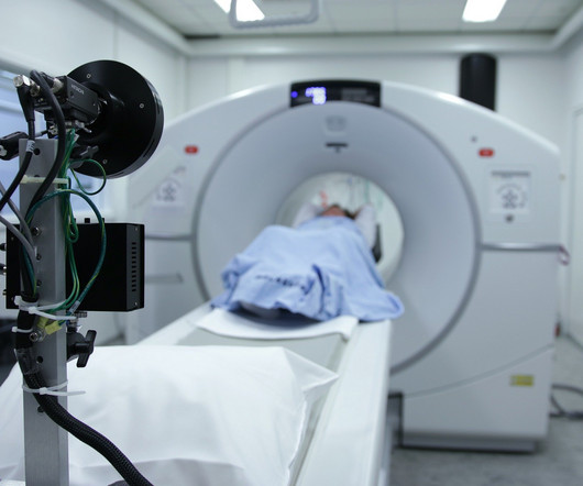
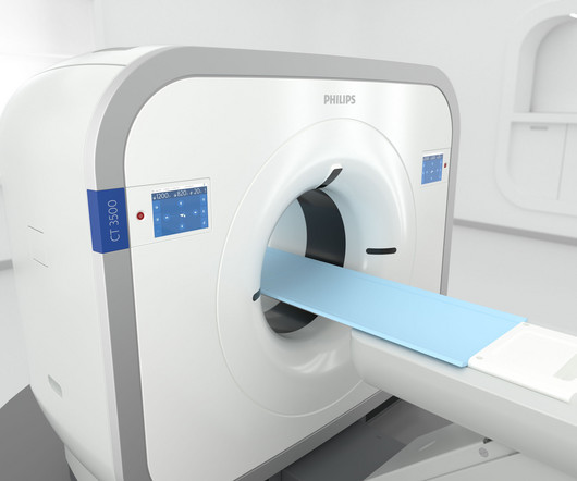
“Increased financial pressures, chronic staff shortages, and escalating patient demand are driving radiology departments to do everything they can to maximize throughput, to guarantee equipment uptime, and to avoid repeat scans,” said Frans Venker, General Manager ComputedTomography at Philips. Results in other cases may vary. [2]
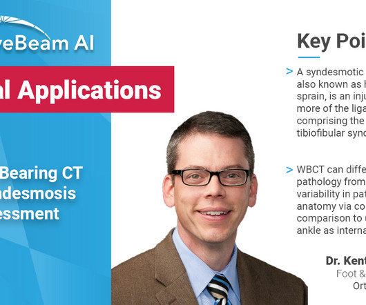
A weight bearing CT scan can: Provide increased sensitivity and specificity over radiographs. Diagnosis If WB X-rays are indeterminate after clinical exam, order a bilateral WBCT. Differentiate pathology from natural variability in patient anatomy via contralateral comparison to uninjured ankle as internal control 1.
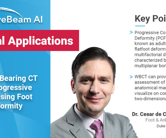
A weight bearing CT scan can: Provide an assessment of important anatomical markers of pronounced hindfoot deformity and peritalar subluxation (PTS), difficult to visualize on conventional two-dimensional radiographs 1. Weightbearing ComputedTomography for Assessment of Foot and Ankle Deformities: The Iowa Experience.
MRI system and a 128- slice computedtomography (CT) system. “At The Essentia SA is an ultra-compact straight arm system, designed for a wide range of standing, sitting and recumbent radiographic exams. To help ease patient tensions, the system features a large 10.5-inch
Common Indications Syndesmosis Provide increased sensitivity and specificity over radiographs 1. Can Weight-Bearing ComputedTomography Be a Game-Changer in the Assessment of Ankle Sprain and Ankle Instability? Cone beam CT of the musculoskeletal system: clinical applications. Help detect subtle syndesmosis injuries 1.
A) AP radiograph of Lisfranc Fracture Dislocation demonstrates the circled “fleck sign” or Lisfranc ligament avulsion fracture fragment. (B) C) The lateral radiograph notes with a circle, the dorsal sub dislocation of the metatarsal base. Radiographs should be repeated after two weeks to ensure surgery is unnecessary.
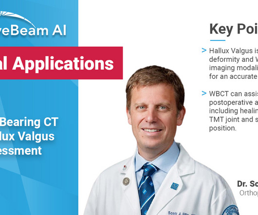
Diagnosis While radiographs are typically sufficient to make the diagnosis, WBCT scans may be useful to plan surgical treatment. Accurately assess sesamoid position as plain radiographs cannot determine whether the sesamoids have been reduced within their grooves 5. . • Assess congruency and degenerative changes at the 1st MTP joint.
KUB indicates kidneys, ureter, and bladder (plain abdominal radiograph); CT, computedtomography; and PCD, percutaneous catheter drainage. In severe cases or patients who do not respond to PCD, treatment with nephrectomy can lead to clinical and radiological improvement (Fig. RadioGraphics. 2000;160(6):797-805.
Frontal abdomen radiograph demonstrates foreign body consistent with capsule endoscopy device (pill cam) in descending colon. Approximately 2% of all capsule endoscopies result in CR [ 2 ] The clinical indication for capsule endoscopy is correlated with different rates of CR. Xray of the Week Figure 1. Gastrointest Endosc.
Computedtomography is also an alternative method for lens subluxation which again can show deviation of the lens (Figs. Radiographics. Sai is also a member of the Gold Humanism Honor Society and is involved with giving back to the community at a local free clinic as a medical assistant. 2006;10(4):345-350.
1 Imaging: Computedtomography (CT) is the recommended imaging modality for evaluating orbital trauma. Clinical features of single and repeated globe rupture after penetrating keratoplasty. Radiographics. Penetrating and blunt trauma, in addition to chemical exposure, account for the majority of cases. Clin Ophthalmol.
They will still play a vital role in advising other doctors which scan would be the most appropriate to answer their clinical query. It is likely that they will lead clinic with patients to discuss their imaging, as already happens in some hospitals. 2011, Radiographics, pp. Monsky, Derek S. Vien, Daniel P. volume 31, issue 5.
Photoprint from radiograph by W.K. 3) In the early twentieth century, it was a common goal for investigators to try to find a way to separate the superimposed shadows that were recorded when a complex structure was shown on a radiograph. (3) 3) This is what is known as tomography. This is now known as ‘Hand mit Ringen’. (1)
When evaluating a CT brain the following, certain cisterns have clinical relevance for potential herniation syndromes, layering of subarachnoid blood, and/or the significant structures that run through them. ComputedTomography. RadioGraphics, 1998; 18(1):151-163 3. In Clinical Neuroanatomy, 28th Edition (2013).
0000000000001232 Cerebral small vessel disease (CSVD) is one of the most common clinical conditions that a neuroimager will encounter. CSVD is associated with an increased risk of clinical ischemic and hemorrhagic stroke, silent infarcts, and cognitive decline and dementia. Continuum (Minneap Minn). 2023;29(2):501-518. doi:10.1212/CON.0000000000001232
Key Points: Imaging modalities such as plain radiographs (X-Ray), computedtomography (CT), and magnetic resonance imaging (MRI), dont have the diagnostic accuracy needed to detect syndesmotic widening or subtle instability.
5,16] Algorithms translate detected changes in MTT, CBV, and CBF into images that can be used in clinical decision-making. Magnetic resonance imaging and computedtomography in emergency assessment of patients with suspected acute stroke: a prospective comparison. In Fitzgerald’s Clinical Neuroanatomy and Neuroscience.
We organize all of the trending information in your field so you don't have to. Join 5,000 users and stay up to date on the latest articles your peers are reading.
You know about us, now we want to get to know you!
Let's personalize your content
Let's get even more personalized
We recognize your account from another site in our network, please click 'Send Email' below to continue with verifying your account and setting a password.


Let's personalize your content