This site uses cookies to improve your experience. To help us insure we adhere to various privacy regulations, please select your country/region of residence. If you do not select a country, we will assume you are from the United States. Select your Cookie Settings or view our Privacy Policy and Terms of Use.
Cookie Settings
Cookies and similar technologies are used on this website for proper function of the website, for tracking performance analytics and for marketing purposes. We and some of our third-party providers may use cookie data for various purposes. Please review the cookie settings below and choose your preference.
Used for the proper function of the website
Used for monitoring website traffic and interactions
Cookie Settings
Cookies and similar technologies are used on this website for proper function of the website, for tracking performance analytics and for marketing purposes. We and some of our third-party providers may use cookie data for various purposes. Please review the cookie settings below and choose your preference.
Strictly Necessary: Used for the proper function of the website
Performance/Analytics: Used for monitoring website traffic and interactions
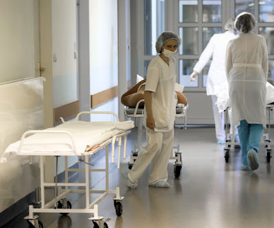
To cover a CT and MR "list" was a luxury; it was to escape the murderous fluoroscopy rooms, intravenous urography (IVU) lists, and indeterminable piles of plain films Then the scanners got fast. So as to report more CT and MRI, radiologists stopped doing hands-on ultrasound and fluoroscopy. 20s rotation, 40s reconstruction.
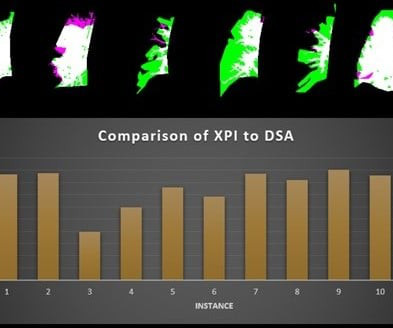
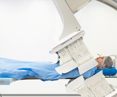
A fluoroscopy method incorporating a noncontrast x-ray pulsatility index (XPI) can improve clinical efficiency as a screening or diagnostic test for suspected chronic thromboembolic pulmonary hypertension, according to research presented at the American Roentgen Ray Society (ARRS) annual meeting.
They found that rates of self-interpretation varied by imaging modality: Self-interpretation rate for office-based imaging by modality Modality Percentage X-ray and fluoroscopy 50.4% First author Eric Christensen, PhD, of HPI and colleagues conducted a study that included data from more than 1.6 Ultrasound 52% Nuclear medicine 39.5%
With interest growing in reevaluating this energy consumption, the researchers equipped seven units at their hospital with power sensors: an interventional radiology suite, a neurointerventional suite, a radiology fluoroscopy unit, two cardiology laboratories, and two urology fluoroscopy units.
VIENNA -- Canon Medical Systems Europe unveiled a new ultrasound scanner and a radiography/fluoroscopy (R/F) system in its booth at ECR 2024. Suitable for clinics as well as large hospitals, Aplio me is a compact (55 x 73 x 130~177 cm) and lightweight system that can be customized to meet the operator’s clinical needs, Canon said.
The team reported the following findings as indicated by respondents: In academic practices, 72% of clinical time was spent in pediatrics compared to 16% in hospital/systems, 5% in private practice, and 4% teleradiology. The respondents represented 376 unique radiology practices across multiple practice settings.
The technology figured prominently in five Minnies categories, including Hottest Clinical Procedure. Pickhardt believes using additional clinical data on CT imaging could save healthcare costs, especially when AI tools are used in parallel, suggesting it is a cost-effective or even cost-saving strategy for personalized or precision medicine.
Clinical use of imaging equipment accounts for more than 50% of greenhouse gas emissions in the radiology department, with MRI and CT equipment major contributors, researchers have reported. Clinical use of imaging equipment across all modalities made up 54% of departmental greenhouse gas emissions (2.5 kt CO 2 e) and CT for 24% (1.1
Radiation Protection Services customers will have access to subject matter expertise in the areas of MRI safety, CT and fluoroscopy dose optimization, and clinical image analysis and review for ACR accreditation.
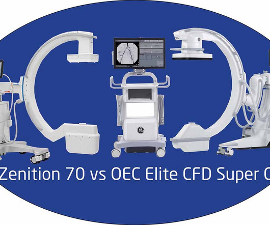
Are you looking for a new C-Arm for your healthcare facility or veterinary clinic? C-Arms use fluoroscopy technology to produce high-resolution real-time X-ray images.
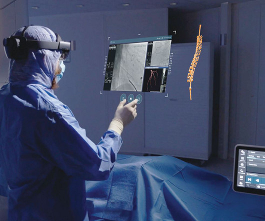
a leading clinical augmented reality med-tech company, announced the successful first installation and clinical cases using the OmnifyXR Interventional Suite [1] at North Star Vascular and Interventional in Minneapolis, Minnesota. Disclaimer: OmnifyXR is intended to be used adjunctively to standard of care imaging.
The clinical utility of DDR is being realized across different provider types, from hospitals to clinics and private practices, encompassing a multitude of applications, including ER/inpatient triage, outpatient imaging, specialty musculoskeletal (MSK) imaging, and pre-surgical evaluations. Zwanger-Pesiri Radiology (N.Y.),
The study, published in the journal Diagnostics, highlights the clinical value of DDR through its unique ability to evaluate diaphragm movement in real time and integrate dynamic functional information with static anatomical data to provide a quantitative assessment of diaphragmatic movement, including excursion and speed.
The new imaging systems that will be on display include three new digital radiography (DR) suites, two new fluoroscopy systems, a 0.4T Persona C-HR: Fujifilm’s newest mobile fluoroscopy c-arm solution providing 30 frames per second FPS pulsed fluoroscopy images at low dose. The sturdy patient table features a robust 800 lbs.
CM stands for the Clinical Modification of the classification system. Category III codes Category III codes are temporary codes that allow for data collection for emerging technologies, services, procedures, and service paradigms. ICD-10 is the 10th edition of this coding system.
The research will produce technical feedback to assist GEHC in assessing the system’s reconstruction methods, image presentation workflow, and clinical benefits for specific pathologies and disease types. MRI GEHC unveiled Signa Champion, a 1.5-tesla, It supports both radiography and fluoroscopic imaging.
Additionally,” said Matthew Smith , MD, from Vanderbilt University Medical Center in Nashville, TN, “this easy-to-implement method can be performed by an x-ray technologist in an outpatient setting,” Smith et al. enrolled volunteers suspected of chronic thromboembolic pulmonary hypertension (CTEPH) based on pulmonary scintigraphy and/or CTA.
The Radiology Experience Tour expanded this year to include a portfolio of Philips imaging solutions including Magnetic Resonance, Radiology and Fluoroscopy, Ultrasound, Radiology Operations Command Center (ROCC), PACS, C-Arms and Ambient Experience. The objectives behind the radiology experience tour are twofold.
The researchers uncovered patterns and implications of abdominal and pelvic injuries in bomb blast victims through radiological and clinical profiling. Fluoroscopy, angiography, and nuclear scans all came in at less than 5%. The team retrieved data from electronic health records. Nearly 30% of the injuries could not be characterized.
The agreement includes hundreds of new systems, including AI-enabled technologies , across nuclear medicine , X-Ray , vascular and cardiovascular ultrasound , regional CT , fluoroscopy, surgery, and bone densitometry for which GE HealthCare will be the sole provider. and Canada.
Fluoroscopy-guided procedures are the leading source of occupational ionizing radiation exposure for electrophysiologic (EP) personnel. Click here to download the full clinical study. Reducing high scatter radiation exposure during medical procedures is the principal task of many professional societies and advisory groups.
CT LVAS, designed for use with computed tomography scans, joins 4D Medical's XV LVAS imaging software cleared for use with fluoroscopy in the United States. As we head to RSNA, I am thrilled that reimbursement has been approved for our XV LVAS product designed for fluoroscopy, and to also share FDA clearance for CT LVAS in the U.S.
Open MRI Equals Greater Comfort, Better Compliance The clinical team at Massac Memorial says the hospital’s Oasis Velocity Open MRI system is boosting efficiency for technologists and offering big benefits to patients. And having that in your own backyard means you’re getting the care you need without having to travel.”
Chapter 3: The Radiologic Toolbox – Types of X-ray Imaging An exploration of the various types of X-ray imaging, including radiography, fluoroscopy, computed tomography (CT), and more. How each modality serves unique clinical purposes and applications.
Chapter 3: Types of X-ray Technology: Beyond Radiography An exploration of the various modalities and applications of X-ray technology, from radiography to fluoroscopy and computed tomography (CT). How each modality serves specific clinical needs and diagnostic challenges.
Chapter 5: Beyond Radiography: The Rise of Advanced Modalities An examination of advanced X-ray modalities, including fluoroscopy, mammography, and computed tomography (CT). How these modalities offer enhanced capabilities for specific clinical applications.
Chapter 3: Types of X-ray Imaging: Beyond Radiography An exploration of the various types of X-ray imaging, including radiography, fluoroscopy, and computed tomography (CT). How each modality is used for different clinical purposes.
Treatment can vary from clinical monitoring, pharmacological therapy, or surgery, depending on the severity and clinical symptoms [4]. The procedure first begins with gaining femoral/jugular vein access and inserting a stiff guidewire into the right atrium, confirmed with fluoroscopy [5,6].
Chest X-Rays should also be used sparingly in COVID-19 suspected individuals and be reserved for those cases where it will impact patient management and is clinically indicated.
UFE is a minimally invasive treatment option where fluoroscopy is used to guide embolic agents to the uterus and fibroids. Sources: The Mayo Clinic John Hopkins Medicine Fibroid Institute Dallas The post THE TRUTH ABOUT UTERINE FIBROIDS appeared first on Delaney Radiology | Wilmington, NC. Uterine Fibroid Embolization (UFE).
Assessment of Claimant, Clinical, and Financial Characteristics of Teleradiology Medical Malpractice Cases. Neuroimaging and Clinical Findings in Healthy Middle-Aged Adults With Mild Traumatic Brain Injury in the PREVENT Dementia Study. Francisco, et al, Emergency Radiology , April 26, 2024. To learn more about this paper, click here.
Greg Nicola “Practices and departments need a subset of radiologists to be on-premise to perform minimally invasive procedures, fluoroscopy, and provide in-person consultation for clinicians," Nicola explained. Greg Nicola, MD, is chair of the American College of Radiology's Commission on Medical Economics.
We organize all of the trending information in your field so you don't have to. Join 5,000 users and stay up to date on the latest articles your peers are reading.
You know about us, now we want to get to know you!
Let's personalize your content
Let's get even more personalized
We recognize your account from another site in our network, please click 'Send Email' below to continue with verifying your account and setting a password.


Let's personalize your content