This site uses cookies to improve your experience. To help us insure we adhere to various privacy regulations, please select your country/region of residence. If you do not select a country, we will assume you are from the United States. Select your Cookie Settings or view our Privacy Policy and Terms of Use.
Cookie Settings
Cookies and similar technologies are used on this website for proper function of the website, for tracking performance analytics and for marketing purposes. We and some of our third-party providers may use cookie data for various purposes. Please review the cookie settings below and choose your preference.
Used for the proper function of the website
Used for monitoring website traffic and interactions
Cookie Settings
Cookies and similar technologies are used on this website for proper function of the website, for tracking performance analytics and for marketing purposes. We and some of our third-party providers may use cookie data for various purposes. Please review the cookie settings below and choose your preference.
Strictly Necessary: Used for the proper function of the website
Performance/Analytics: Used for monitoring website traffic and interactions
. | S1-SSCH01-5 | E451A This scientific paper may increase overall confidence in the potential of using multimodal AI for tuberculosis (TB) detection, and potentially autonomous reporting, on chest radiographs in certain clinical settings. Stop by the session with your questions.
Some of my radiological heroes would report a staggering 30,000 to 40,000 radiographs a year. Some even [startled gasp] gave up reporting plain radiographs. But the pressure kept building, and the number of CT and MRI scans grew by 20% annually in my hospital. I still don’t know how they did it. It is unsafe for our patients.
"Practical AI implementation will require objective onsite performance evaluation, institutional information technology infrastructure integration, and postdeployment monitoring," wrote a team led by Eui Jin Hwang, MD, PhD, of Seoul National University Hospital in South Korea. van Beek, MD, of the University of Edinburgh in the U.K.
Middlebrooks, MD, Mayo Clinic, Jacksonville, FL Erik H. Middlebrooks, MD, of the Mayo Clinic in Jacksonville, FL. Given the extensive array of sequences and techniques employed in clinical imaging, we must address the inherent challenges associated with each," he noted. I'm a radiographer,' " Stewart recalled.
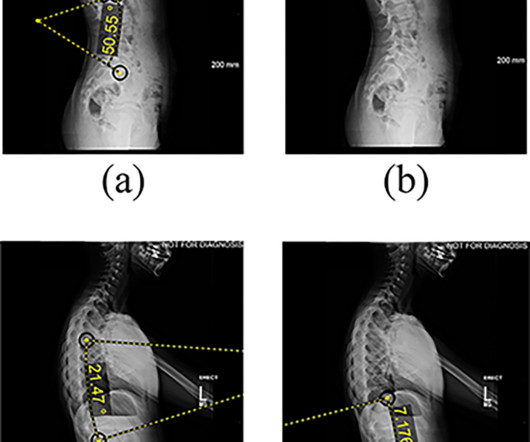
Chest dynamic digital radiography (DDR) may have received a boost toward clinical use in patients with lung disorders, with researchers developing AI to perform time-consuming analysis involved in the technology, according to researchers in New York City. a) Raw example of a dynamic digital radiograph. (b)
The finding by researchers in Copenhagen, Denmark, suggests that AI could eventually help streamline high-volume radiology workflows by handling some of the more “tedious parts of the work,” lead author Louis Plesner, MD, of Herlev and Gentofte Hospital in Denmark told AuntMinnie.com. The full study can be found here.
Most Influential Radiology Researcher Erik Middlebrooks, MD, Mayo Clinic, Jacksonville, FL Erik Middlebrooks, MD. One of this year's nominations for Most Influential Radiology Researcher is Erik Middlebrooks, MD, of the Mayo Clinic in Jacksonville, FL. He earned his medical degree from Washington University in St.
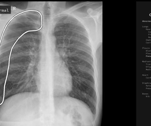
Gleamer) increased sensitivity for detecting all abnormalities on x-rays for all readers (thoracic radiologists, general radiologists, and radiology residents), according to a group of Gleamer consultants and clinicians at Cochin Hospital in Paris. In a retrospective study, a commercially available algorithm (ChestView, v.
tesla MRI AI body composition analysis Cardiac PET Cryo/thermoablation CT colonography Genicular artery embolization Hyperpolarized xenon-129 MRI PET/MRI Photon-counting CT Radiomics Theranostics Whole-body MRI screening Image of the Year 3D PET/MR image.
Deep-learning models could have potential as predictive tools for breast cancer prognosis, a study published January 17 in Clinical Breast Cancer has found. From there, the team used clinical data and imaging characteristics to select independent prognostic factors to establish a clinical model.
Charlene Liew, MD, director of cardiothoracic imaging at Changi General Hospital in Singapore, outlined the current state of AI in radiology and where it is going in a presentation called "AI in Radiology: The Past Informs the Future."
The finding validates fully automated software for use in procedures and advances DDR a step toward clinical use, the technology’s U.S. Representative chest radiographs and graphs of lung signal intensity obtained using DDR for the severe COPD group. and Japanese developers wrote. The subject is a 77-year-old male with VC of 1.68
It's a question researchers at University College London Hospitals NHS Foundation Trust and Canterbury Christ Church University have asked as part of their prospective, randomized, multisite trial currently open and underway in the U.K. How does immediate AI-enabled patient triage on chest CT impact the lung cancer pathway?
Presenting the research on November 28 at RSNA 2023, Jan Rudolph, MD, from the department of radiology at University Hospital LMU Munich said nonradiologists can significantly benefit from AI assistance in emergency-related chest x-ray analysis. “If
. | W3-SSMK08-4 | Room E450A A deep learning-based framework for automated screening of osteoporosis on lumbar spine plain radiographs shows potential as another way to opportunistically make use of imaging studies performed for other indications, according to this presentation.
A team led by Yin Ting Chiu, PhD, from the Hong Kong Children’s Hospital discussed key factors identified by the hospital’s MR team for effectively performing supplementary MRI scans on children ages three to seven without sedation.
The current model has potential to serve as a valuable clinical tool, providing insight into the optimal timing of intervention and surgical planning parameters,” the group wrote. Features extracted from lateral radiographs. (a) AVBT was approved in the U.S. The full study can be found here.
In this AJR accepted manuscript , a DL model was developed in 7,105 patients via one institution from March 2013 to December 2019 (3:1:1 allocation to training, validation, and internal test sets) to predict risk of all-cause mortality within 30 days after CAP diagnosis using patients’ initial chest radiograph. CURB-65 score). “The
The technology figured prominently in five Minnies categories, including Hottest Clinical Procedure. Pickhardt believes using additional clinical data on CT imaging could save healthcare costs, especially when AI tools are used in parallel, suggesting it is a cost-effective or even cost-saving strategy for personalized or precision medicine.
In an open forum, Yi Xiang Tay, of Singapore University Hospital's radiography and diagnostic imaging department, shared his team's research. In addition, clinical decision support systems were the most evaluated mode of intervention, either integrated or standalone. For more coverage from ECR 2024, please visit our RADCast.
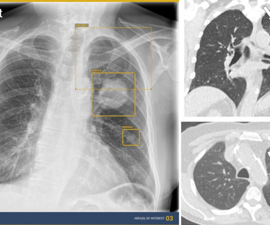
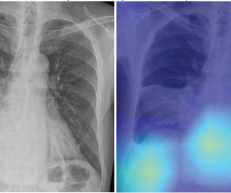
This competition demonstrated the value of AI in detecting and localizing many pathologies in chest radiographs by simulating the real work situations of radiologists,” the group wrote. The study was published February 8 in the Journal of Imaging Informatics in Medicine. The full study is available here.
“This is a potentially practice-changing trial,” said Oliver Sartor, MD, a medical oncologist and director of radiopharmaceutical trials at the Mayo Clinic in Rochester, MN. He presented the abstract on behalf of first author Ken Herrmann, MD, chair of nuclear medicine at the University Hospital Essen in Essen, Germany.
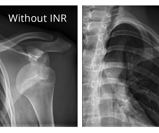
The company will showcase the clinical analysis of Canon’s Intelligent Noise Reduction (Intelligent NR) that provides superior image quality while lowering radiation dosing in pediatric digital radiography at the Radiological Society of North America Annual Meeting 2023 (RSNA) , McCormick Place Convention Center, Chicago, IL Nov.
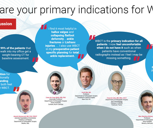
The following panelists of experts share their insights and experiences in the use of WBCT in clinical practice as well as the future direction of the modality. Whenever bilateral standing radiographs would have been needed, a WBCT was performed instead. How Frequently Do You Use It In Practice?
Reading Time: 10 minutes read By Henry Williams, Carestream Area Vice President, Sales Western Nowadays, with hospital budgetary restrictions at the forefront of the purchasing decision making process, it seems like the X-Ray market, like everything else, is not immune to the current state of the economy. Who is Making the Purchases?
When the radiology department at Cork University Hospital in the south coast of Ireland took the lead in developing an autism-friendly patient experience, they observed that it took less time to complete imaging studies for children with autism, according to a February 28 session at ECR 2024.
meets this need by developing clinical-grade AI models designed to improve capacity. solutions are driving a significant improvement through early lung cancer detection within hospital systems. Averaged across all findings on chest radiographs.) [2]AIDE Co-Founder and Chief Executive Officer. million members. Published 2021.
S1-SSCH01-5 | E451A This scientific paper may increase overall confidence in the potential of using multimodal AI for tuberculosis (TB) detection, and potentially autonomous reporting, on chest radiographs in certain clinical settings. First PANORAMA findings support AI-based opportunistic screening Sunday, December 1 | 10:40 a.m.-10:50
Knee osteoarthritis (OA) clinical trials results show that weight bearing CT (WBCT) imaging may offer new insights into OA pathology beyond what plain radiographs and MRI can provide. Some of these findings could immediately be translated into clinical practice as well. of medial and 8.5% of medial and 8.8% of lateral menisci.
Key Points: Currently plain radiographs are the standard method in diagnosing syndesmotic ankle injuries even though the distal tibiofibular joint cannot be assessed due to superposition of the osseous structures in the foot. Dr. Peiffer et. Researchers used data from 76 NWBCT ankles (retroactive study) and 86 WBCT ankles.
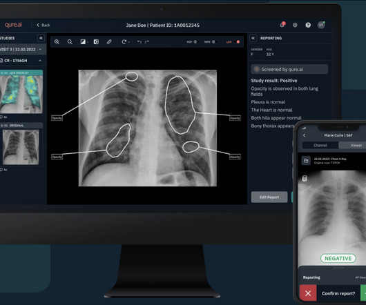
Chest radiography is the most common medical imaging tool used in routine clinical practices to identify different disease findings. The company's products are deployed in over 500 hospitals and imaging centers, impacting the lives of over a million individuals every year. billion annual X-rays performed, 1.5
Researchers found that first-ray hypermobility primarily arises from TMT1 instability, though there is still a limited understanding of TMT1 morphology and anatomy due to the challenges and limitations of radiographic imaging. To read the full study click here.
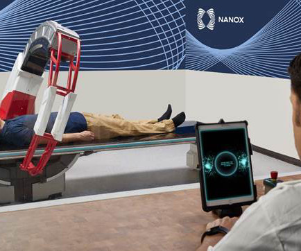
Following this clearance, Nanox will continue to work with the FDA to pursue additional regulatory clearances and intends to expand clinical indications. regulatory clearance also paves the way for Nanox.ARC to be approved in other countries that are FDA-clearance-based markets. While access to medical imaging is relatively high in the U.S.
In the third blog of her series on AI and the radiographer, Shamie Kumar explores the impact on the radiographer when AI is integrated within an imaging modality. The question to explore in this blog is when AI is integrated within an imaging modality itself and how that may impact a radiographer.
Specializing in abdominal radiology, Dr. Lee is chief of women’s imaging and officer of mentored research in the Department of Radiology at Massachusetts General Hospital ( MGH ) and associate professor of radiology at Harvard Medical School in Boston. “I The journal will be published in partnership with Oxford University Press.
Reading Time: 8 minutes read By Dr. Niall Sheehy, MB, MRCP, FFR RCSI, St James’s Hospital -Trinity College Dublin. In May 2021, St James’s Hospital in Dublin was one of 54 public hospitals affected when the Health Service Executive (HSE) was the victim of a Cyber Attack by Conti, a sophisticated, financially-motivated criminal gang.
The following panelists of experts share their insights and experiences in the use of WBCT in clinical practice as well as the future direction of the modality. Therefore, its indications are the same as radiographs. I no longer refer patients for radiographs or conventional CT in any case. Even soft tissue issues (i.e.
It can also serve as a crucial second reader for physicians, assisting in the review of frontal (AP/PA) chest radiographs of adults acquired on digital radiographic systems. In the second pivotal study, a landmark clinical evaluation of qXR-LN was conducted through a multi-reader, multi-case clinical validation study.
While clinical seizures are readily identifiable, subclinical seizures—which comprise most seizures after a hemorrhage—are hard to detect. Thereafter, clinical seizures were objectively reported. OUTCOMES Primary: Occurrence of at least one clinical or electrographic seizure recorded on continuous EEG within 72 hours after inclusion.
Reading Time: 9 minutes read Mid-cycle refresh can increase clinical, operational, and security benefits. Does it support new medical imaging software that can help improve clinical outcomes? Physicians have better diagnostic confidence when they can view radiographs in the manner most suitable to their preferences.
PMID: 36693146 Clinical Question: In patients aged 18-50 years with primary and complete pneumothorax, is simple aspiration non-inferior to chest tube drainage for rates of pulmonary re-expansion within 24 hours? Article: Marx T, Joly LM, Parmentier AL, et al. Am J Respir Crit Care Med. 2023;207(11):1475-1485.
PMID: 37188358 Clinical Question: What is the efficacy and safety of low-dose (25mg) prolonged administration (over 6hrs) of alteplase in patients with massive PE? Click here for Direct Download of the Podcast Paper: Aykan AC et al. Reduced-Dose Systemic Fibrinolysis in Massive Pulmonary Embolism: A Pilot Study. Clin Exp Emerg Med 2023.
But how will AI in the workplace affect the radiographer and how does it differ from the red dot system radiographers are so familiar with? The Red Dot System Often one of the first courses a newly qualified radiographer attends is the red dot course. What does AI do that a radiographer doesn’t already?
Kim Mason Kim Mason, an Audit and Research Radiographer for Mid Yorkshire Teaching Hospitals Trust, talks about their role as well as the value of radiographer engagement in research activities and how to get involved. So, what is an Audit and Research Radiographer? However, I’m not entirely non-clinical.
We organize all of the trending information in your field so you don't have to. Join 5,000 users and stay up to date on the latest articles your peers are reading.
You know about us, now we want to get to know you!
Let's personalize your content
Let's get even more personalized
We recognize your account from another site in our network, please click 'Send Email' below to continue with verifying your account and setting a password.


Let's personalize your content