This site uses cookies to improve your experience. To help us insure we adhere to various privacy regulations, please select your country/region of residence. If you do not select a country, we will assume you are from the United States. Select your Cookie Settings or view our Privacy Policy and Terms of Use.
Cookie Settings
Cookies and similar technologies are used on this website for proper function of the website, for tracking performance analytics and for marketing purposes. We and some of our third-party providers may use cookie data for various purposes. Please review the cookie settings below and choose your preference.
Used for the proper function of the website
Used for monitoring website traffic and interactions
Cookie Settings
Cookies and similar technologies are used on this website for proper function of the website, for tracking performance analytics and for marketing purposes. We and some of our third-party providers may use cookie data for various purposes. Please review the cookie settings below and choose your preference.
Strictly Necessary: Used for the proper function of the website
Performance/Analytics: Used for monitoring website traffic and interactions
Intelligent virtual and AI-based collimation features appear to save radiographers time during x-ray image acquisitions – a key function for enabling more patient-focused workflows, according to a recent study. To that end, the researchers conducted an observational study at five clinical sites in Europe and the U.S.
The use of AI in thoracic imaging has begun to demonstrate "cumulative evidence of effectiveness," but more testing and research are needed to determine its feasibility for this application, according to a commentary published February 25 in Radiology. van Beek, MD, of the University of Edinburgh in the U.K. in an accompanying editorial.
In a reader study involving five radiologists interpreting 758 chest x-rays, use of the model (AIRead, Soombit.ai) reduced average reading times by 14 seconds per image and increased sensitivities for certain findings. Image and caption courtesy of RSNA. AP = anterior-posterior. seconds 20.4 seconds 12.5
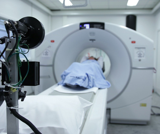
Some of my radiological heroes would report a staggering 30,000 to 40,000 radiographs a year. The scanners could reconstruct images faster than we could report them. Some even [startled gasp] gave up reporting plain radiographs. A good day five to 10 years ago was reporting 40 cross-sectional imaging studies.
Middlebrooks, MD, Mayo Clinic, Jacksonville, FL Erik H. Middlebrooks, MD, of the Mayo Clinic in Jacksonville, FL. His lab team is currently focused on "implementing advanced techniques, such as parallel transmit, to achieve more consistent image quality and fully harness the power of 7T for every patient," Middlebrooks said.
Chest dynamic digital radiography (DDR) may have received a boost toward clinical use in patients with lung disorders, with researchers developing AI to perform time-consuming analysis involved in the technology, according to researchers in New York City. a) Raw example of a dynamic digital radiograph. (b)
Deep-learning models could have potential as predictive tools for breast cancer prognosis, a study published January 17 in Clinical Breast Cancer has found. They collected imaging data to establish deep-learning models using ResNet50. This means the combined model could predict prognosis after surgery.
The finding validates fully automated software for use in procedures and advances DDR a step toward clinical use, the technology’s U.S. DDR is a novel functional imaging technique that uses sequential images obtained by a pulsed x-ray generator and a flat panel detector with a large field of view. and Japanese developers wrote.
The new rules now in effect in Michigan help ensure that only medical radiologic technologists with the appropriate education and training can operate medical imaging equipment, according to an American Registry of Radiologic Technologists (ARRT) legislative update. Both the ARRT and the MSRT are opposing that measure.
These included chest radiographs that displayed abnormalities of no clinical significance, which are typically treated as normal. Reports by radiologists for the images were classified similarly. of unremarkable chest radiographs, while only missing 0.1%, 1%, and 2% of remarkable chest radiographs.
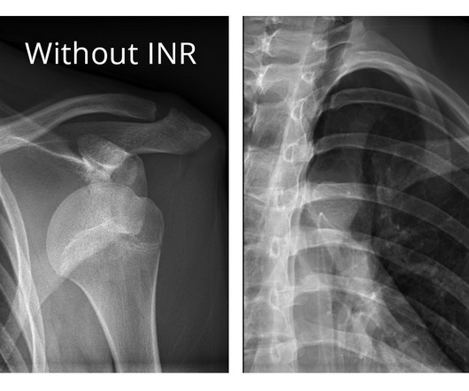
Researchers at the Mayo Clinic in Rochester, MN, have taken a first step in using AI to automatically classify and organize shoulder x-rays on a large scale, according to a recent study. To date, most registries consist primarily of clinical data, and either completely lack or contain very limited medical imaging information.
Implementing imaging referral guidelines not only supports value-based radiology but makes it easier to communicate with patients about low-value services, according to findings presented February 29 at ECR 2024. Low-value imaging procedures result in an ineffective allocation of resources and imposes potential danger," Tay said.
Are low-field MRI units effective for neuroradiologic imaging? The group's commentary was published February 13 in RadioGraphics. The group's commentary was published February 13 in RadioGraphics. It's true that overall image quality of routine clinical brain and spinal MR imaging at 0.55-tesla
VIENNA -- It's definitely possible to develop a successful cardiac imaging practice, according to a professional development presentation delivered February 28 at the ECR in Vienna. Gutberlet described how he came to cardiac imaging early in his medical career. years of follow-up compared with CT imaging (2.1% But he persevered.
The need for imaging modalities to support an earlier, more accurate diagnosis continues to drive AI applications in the medical imaging market. In fact, the size of the market for AI in medical imaging is experiencing phenomenal growth, expected to increase from $1.12 billion in 2022 to $27.52
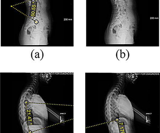
An AI model has delivered a long overdue update of pediatric bone growth predictions used in x-ray imaging to monitor scoliosis, according to a study published April 30 in Radiology. Full-body biplanar slot scanning is a type of low-dose digital x-ray imaging used to monitor scoliosis. Image courtesy of Radiology.
Most Influential Radiology Researcher Erik Middlebrooks, MD, Mayo Clinic, Jacksonville, FL Erik Middlebrooks, MD. One of this year's nominations for Most Influential Radiology Researcher is Erik Middlebrooks, MD, of the Mayo Clinic in Jacksonville, FL. There's a huge role for AI in image acquisition," he said.
Of course, AI also continues to advance in breast imaging, and we’ll provide coverage of key sessions in our upcoming Women’s Imaging section of the Road to RSNA. AI highlights clinically significant prostate cancer on MRI Sunday, November 26 | 9:30 a.m.-9:40 9:40 a.m. | 2:00 p.m. | Sunday, November 26 | 3:20 p.m.-3:30
ChatGPT-4 outperformed human clinicians in determining pretest and post-test disease probability after a negative test result involving chest radiographs and mammograms, according to a research letter published December 11 in JAMA Network Open. They compared its performance with a survey of 553 human clinicians from various specialties.
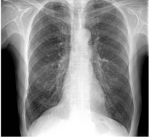
Our findings suggest that artificial intelligence assistance in chest radiograph interpretation may enhance sensitivity without affecting specificity for all readers, regardless of their level of expertise or seniority or the type of abnormality,” wrote first author Souhail Bennani, MD, and colleagues. Image courtesy of Radiology.
SINGAPORE - AI is already changing the practice of medical imaging, and there's more technological growth to come, according to a plenary talk delivered May 4 at the International Society of Magnetic Resonance in Medicine (ISMRM) annual meeting. Food and Drug Administration (FDA) were for radiology indications.
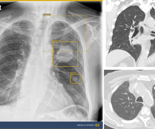
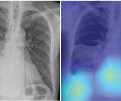
Clinical centers rarely have the necessary resources and personnel to evaluate and compare multiple products prior to purchase,” the group wrote. A) Radiograph in a man (age, 72 years) with a nodule present (reference standard score, 100) shows a true-positive result based on the average algorithm scores. (B)
The technology figured prominently in five Minnies categories, including Hottest Clinical Procedure. Using understandable AI, OSCAR will connect imaging studies from 10 or more years ago to downstream events, Pickhardt explained. His work on abdominal imaging has led to over 500 scientific publications. And 2023 is no exception.
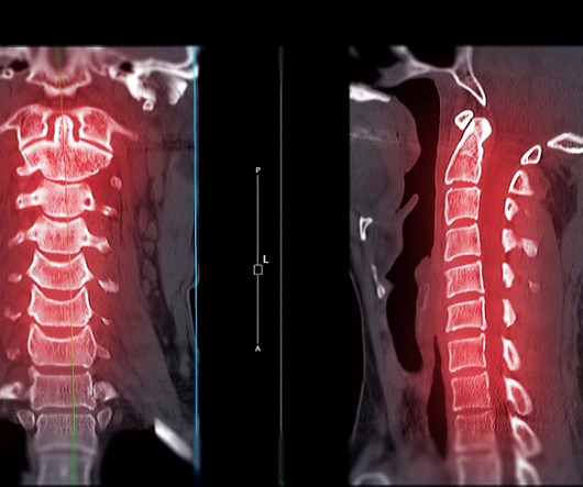
A highly accurate prediction rule for cervical spine injuries in children reduces the use of CT scans by over 50% without missing clinically significant injuries or increasing the use of unnecessary X-rays.
. | W3-SSMK08-4 | Room E450A A deep learning-based framework for automated screening of osteoporosis on lumbar spine plain radiographs shows potential as another way to opportunistically make use of imaging studies performed for other indications, according to this presentation.
An AI model for x-ray imaging could help clinicians plan treatment other than spinal fusions in patients with adolescent idiopathic scoliosis, according to research published January 14 in PLOS One. Features extracted from lateral radiographs. (a) Image courtesy of PLOS One. AVBT was approved in the U.S.
This competition demonstrated the value of AI in detecting and localizing many pathologies in chest radiographs by simulating the real work situations of radiologists,” the group wrote. The study was published February 8 in the Journal of Imaging Informatics in Medicine. Image courtesy of the Journal of Imaging Informatics in Medicine.
Ultimately, the group described a method to correct the phenomenon to produce better images. The article was published March 7 in IEEE Transactions on Medical Imaging. This ensures an unobstructed qualitative evaluation, and enables a quantitative evaluation of dark-field radiographs,” Urban et al wrote.
The deep learning (DL) model may guide clinical decision-making in the management of patients with CAP by identifying high-risk patients who warrant hospitalization and intensive treatment,” concluded first author Eui Jin Hwang, MD, PhD, from the department of radiology at Seoul National University College of Medicine in Korea. Hwang et al.
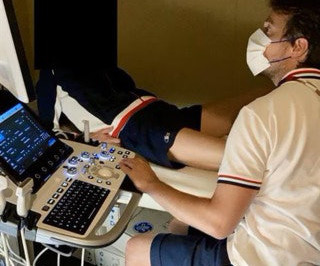
A team of 32 radiologists and 36 radiographers are limbering up to work at the summer Olympics, which begins July 25. Clinical information is essential to refine the diagnosis, but we must also understand the sporting issues in order to adapt the athletes’ care.” Image courtesy of Michel Daoud Crema, MD. “I
“Although dynamic visualization of anatomy is useful to detect numerous pathologies, quantitative assessment of joint motion may improve sensitivity and specificity of DRR,” Sabol said, in a scientific session on musculoskeletal imaging. These images are then processed to visualize joints in motion.
Radiologists can help reduce unnecessary follow-up work in patients with suspected oropharynx cancer by analyzing certain metrics on PET/CT scans, according to a team of head and neck surgeons at the Mayo Clinic in Rochester, MN.
Communication and engagement with child-life specialists are a couple of factors in successful MR imaging for young children, according to research presented May 4 at the International Society for Magnetic Resonance in Medicine (ISMRM) meeting in Singapore. The team also added that the parent-child combo is also useful.
MRI helps clinicians assess the neural involvement in endometriosis and could help them prevent irreversible nerve damage and chronic sensorimotor neuropathy in women suffering from the condition, Cleveland Clinic researchers have reported. The team's review of MRI's role for this indication was published January 3 in RadioGraphics.
The company will showcase the clinical analysis of Canon’s Intelligent Noise Reduction (Intelligent NR) that provides superior image quality while lowering radiation dosing in pediatric digital radiography at the Radiological Society of North America Annual Meeting 2023 (RSNA) , McCormick Place Convention Center, Chicago, IL Nov.
This is a potentially practice-changing trial,” said Oliver Sartor, MD, a medical oncologist and director of radiopharmaceutical trials at the Mayo Clinic in Rochester, MN. Radiographic progression-free survival (rPFS) was the primary endpoint of the study, while overall survival was the key secondary endpoint.
The study, published in the journal Diagnostics, highlights the clinical value of DDR through its unique ability to evaluate diaphragm movement in real time and integrate dynamic functional information with static anatomical data to provide a quantitative assessment of diaphragmatic movement, including excursion and speed.
milla1cf Fri, 07/07/2023 - 21:32 July 7, 2023 — FUJIFILM Healthcare Americas Corporation, a leading provider of diagnostic and enterprise imaging solutions, today announced the U.S. X-rays are the most widely used diagnostic tests, accounting for 60% of all imaging studies conducted.
Weight bearing CT (WBCT) imaging has fundamentally changed the evaluation and management of foot and ankle disorders such as hallux valgus, progressive collapsing foot disorder (PCFD), ankle instability, and traumatic deformities. Whenever bilateral standing radiographs would have been needed, a WBCT was performed instead.
AI algorithms appear to have clinical value based on detecting normal x-rays – that is, by flagging chest x-rays as normal versus abnormal, they may reduce reading times for radiologists, according to research presented recently at the RSNA meeting in Chicago. In a session on chest imaging, scientists from AI developers Lunit and DeepTek.ai
Detecting CSIs in a clinical setting often requires imaging such as X-rays and computed tomography (CT) scans, both of which expose children to radiation, which can cause other health issues over time.
Shamie Kumar describes how AI fits into a radiology clinical workflow and her perspective on how a clinicalradiographer could use this to learn from and enhance their skills. If the AI findings are seen in PACS, how many radiographers actually log into PACS after taking a scan or X-ray? Can Radiographers Up-Skill?
"Lack of AI knowledge among educators was the top reason for not integrating AI in education," noted a team led by MRI radiographer Nikolaos Stogiannos of the University of London in the U.K. The study was published July 13 in the Journal of Medical Imaging and Radiation Sciences. the authors concluded.
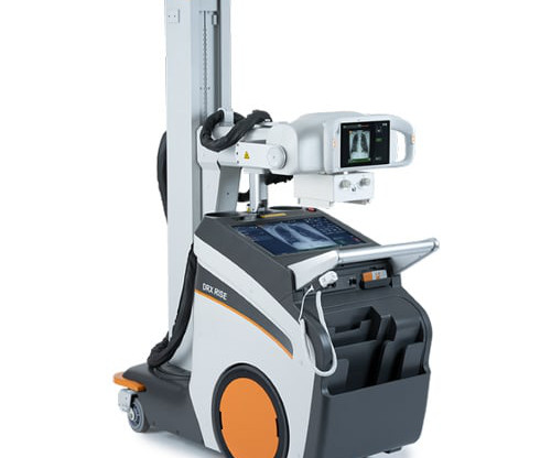
Carestream’s new solutions that will be at AHRA: DRX-Rise Mobile X-ray System — a new, fully integrated mobile X-ray unit packed with efficiency-boosting features, giving customers an affordable path to digital imaging; the DRX-Rise provides optimal-quality images while increasing throughput.
We organize all of the trending information in your field so you don't have to. Join 5,000 users and stay up to date on the latest articles your peers are reading.
You know about us, now we want to get to know you!
Let's personalize your content
Let's get even more personalized
We recognize your account from another site in our network, please click 'Send Email' below to continue with verifying your account and setting a password.


Let's personalize your content