This site uses cookies to improve your experience. To help us insure we adhere to various privacy regulations, please select your country/region of residence. If you do not select a country, we will assume you are from the United States. Select your Cookie Settings or view our Privacy Policy and Terms of Use.
Cookie Settings
Cookies and similar technologies are used on this website for proper function of the website, for tracking performance analytics and for marketing purposes. We and some of our third-party providers may use cookie data for various purposes. Please review the cookie settings below and choose your preference.
Used for the proper function of the website
Used for monitoring website traffic and interactions
Cookie Settings
Cookies and similar technologies are used on this website for proper function of the website, for tracking performance analytics and for marketing purposes. We and some of our third-party providers may use cookie data for various purposes. Please review the cookie settings below and choose your preference.
Strictly Necessary: Used for the proper function of the website
Performance/Analytics: Used for monitoring website traffic and interactions
Chest dynamic digital radiography (DDR) may have received a boost toward clinical use in patients with lung disorders, with researchers developing AI to perform time-consuming analysis involved in the technology, according to researchers in New York City. a) Raw example of a dynamic digital radiograph. (b)
Staff operating ionizing radiation equipment in Michigan are now required to meet certain qualifications for active status and employment. The rules also establish initial and continuing education requirements for limited-scope radiographers and radiologist assistants. Read the full filing here.
Most Influential Radiology Researcher Erik Middlebrooks, MD, Mayo Clinic, Jacksonville, FL Erik Middlebrooks, MD. One of this year's nominations for Most Influential Radiology Researcher is Erik Middlebrooks, MD, of the Mayo Clinic in Jacksonville, FL.
At RSNA 2023, look for AI-driven systems that radiographers can use to help make patient positioning faster and more precise, and bring consistency to the process, all of which help improve image quality and reduce the need for retakes. Even the most skilled radiographers can fail to get positioning just right.
"Lack of AI knowledge among educators was the top reason for not integrating AI in education," noted a team led by MRI radiographer Nikolaos Stogiannos of the University of London in the U.K. The study was published July 13 in the Journal of Medical Imaging and Radiation Sciences. the authors concluded.
DDR is an emerging imaging technique that uses a pulsed x-ray source to acquire a series of radiographs at six to 15 frames per second. These images are then processed to visualize joints in motion.
Communication among practitioners: Effective communication between radiographers, radiologists, and child-life specialists is needed to discuss scan protocols, streamline the exam, and minimize table time. Having support from a child-life specialist inside the scan room can help children stay calm and cooperative.
“So we were thinking and asking ourselves, ‘can nonradiologists benefit from AI and chest radiography analysis in this emergency unit set.’ ” Per year, LMU receives between 5,000 and 6,000 orders for chest radiographs for primary diagnosis from the emergency unit alone.
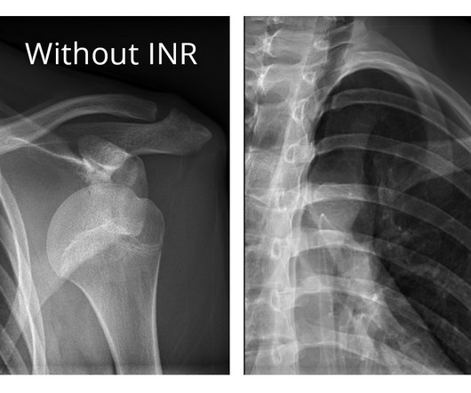
The company will showcase the clinical analysis of Canon’s Intelligent Noise Reduction (Intelligent NR) that provides superior image quality while lowering radiation dosing in pediatric digital radiography at the Radiological Society of North America Annual Meeting 2023 (RSNA) , McCormick Place Convention Center, Chicago, IL Nov.
Tay said while radiology professional societies have advocated incorporating evidence-based imaging referral guidelines into clinical practice, the extent of progress in implementing these guidelines varies worldwide. In addition, clinical decision support systems were the most evaluated mode of intervention, either integrated or standalone.
The study, published in the journal Diagnostics, highlights the clinical value of DDR through its unique ability to evaluate diaphragm movement in real time and integrate dynamic functional information with static anatomical data to provide a quantitative assessment of diaphragmatic movement, including excursion and speed.
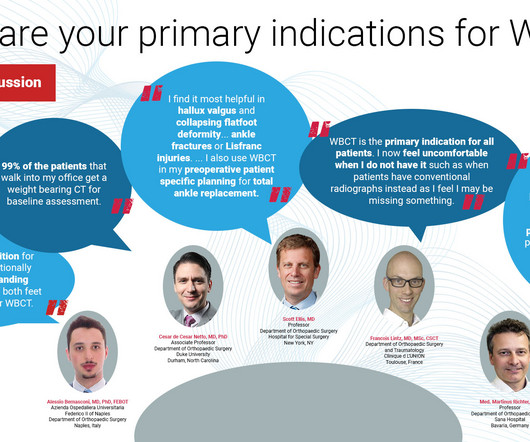
The following panelists of experts share their insights and experiences in the use of WBCT in clinical practice as well as the future direction of the modality. Whenever bilateral standing radiographs would have been needed, a WBCT was performed instead. How Frequently Do You Use It In Practice?
Shamie Kumar describes how AI fits into a radiology clinical workflow and her perspective on how a clinicalradiographer could use this to learn from and enhance their skills. If the AI findings are seen in PACS, how many radiographers actually log into PACS after taking a scan or X-ray? Can Radiographers Up-Skill?
Detecting CSIs in a clinical setting often requires imaging such as X-rays and computed tomography (CT) scans, both of which expose children to radiation, which can cause other health issues over time.
The ASRT weighed in, telling AuntMinnie.com, "A key issue we continue to face is state legislation designed to weaken or remove licensure for medical imaging and radiation therapy professionals. For CT and MRI procedures where injected contrast media is ordered, ".
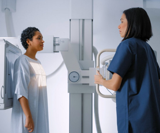
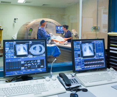
Reading Time: 9 minutes read Carestream solutions help improve patient safety and clinical outcomes. A fundamental goal of radiographers is to complete an imaging exam that provides sufficient information for an accurate clinical diagnosis–and at the lowest possible dose.
Knee osteoarthritis (OA) clinical trials results show that weight bearing CT (WBCT) imaging may offer new insights into OA pathology beyond what plain radiographs and MRI can provide. Some of these findings could immediately be translated into clinical practice as well. of medial and 8.5% of medial and 8.8% of lateral menisci.
Dynamic chest radiography (DCR) shows potential as a tool to investigate lung health in people with cystic fibrosis (CF), according to research published February 13 in Clinical Radiology. and colleagues. Yet FEV1 may not always reflect the severity of the airway obstruction, they noted.
CBCT is an innovative imaging modality with a body of supporting literature reporting reduced radiation exposure, shortened examination time and operating time, and a decreased time interval between injury and diagnosis. Researchers analyzed the limited literature available on using CBCT as a primary modality for diagnosing AS and CLAI.
Research demonstrates that ultrasound substantially enhances the detection of clinically significant, small, mostly invasive, and node-negative cancers. The shortage of radiographers: A global crisis in healthcare. J Med Imaging Radiat Sci. 2023 Oct 19:S1939-8654(23)01877-5. doi: 10.1016/j.jmir.2023.10.001. 2023.10.001.
Dr. Lee’s clinical and scientific interests include abdominal and pelvic imaging, women’s imaging , radiation safety, advanced MRI and CT , molecular imaging and gynecological cancers. Lee has served as a principal investigator in several National Cancer Institute (NCI)-funded clinical trials.
It automates radiographers’ most time-consuming steps so that they can spend more time focusing on the patient.” Precise Image allows radiology departments to simultaneously achieve up to 60% improved low-contrast detectability, 85% lower noise, and 80% lower radiation dose. Results in other cases may vary. [2]
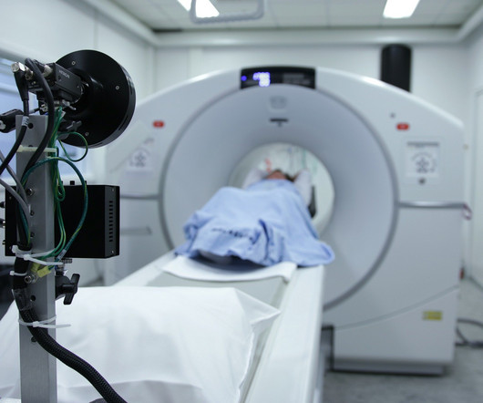
Not only are children more radiosensitive than adults (the cancer risk per unit dose of ionizing radiation is higher), but children also have a longer expected lifetime, which puts them at greater risk of cancer following radiation exposure.(1) The balance of dose and image quality is even more important in pediatric medical imaging.
The following panelists of experts share their insights and experiences in the use of WBCT in clinical practice as well as the future direction of the modality. Therefore, its indications are the same as radiographs. You just get a CT as a bonus, without extra radiation, and it is much faster. Even soft tissue issues (i.e.
Kim Mason Kim Mason, an Audit and Research Radiographer for Mid Yorkshire Teaching Hospitals Trust, talks about their role as well as the value of radiographer engagement in research activities and how to get involved. Hi, I’m Kim and I am an alternative-styled, funky-haired, septum-pierced, disabled Audit and Research Radiographer.
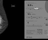
Transforming the breast dose model The prestigious Joint AAPM Task Group 282/EFOMP Working Group, focused on modernizing breast radiation dose modeling, recently published a report outlining their new model in Medical Physics. Volumetric breast density emerges as an essential input to facilitate patient-based radiation dose estimates.
Radiation oncology clinics face numerous challenges in the present environment, including the simultaneous management of multiple tasks (many of which are manual in nature), various degrees of standardisation, and the potential for errors to impact patient treatment. Healthcare Quarterly. April 2012. doi:10.12927/hcq.2012.22845
How are radiographers treated when they admit to making a mistake? And are clinical staff treated the same across disciplines when they make mistakes? Incorrect markers can result in a patient having to undergo a second exam and additional exposure to radiation. Some safety incidents have nothing to do with radiation.
Often an “error” is determined later in the light of additional information and a developing clinical picture. Correction: initially review the study blindly before reading the clinical information. But I strongly believe that these activities are essential to improving our clinical practice. Radiographics (2015);35:1668-1676.
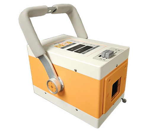
These lightweight and easily transportable units empower veterinarians to conduct imaging procedures right in the comfort of the animal’s familiar surroundings, whether that be in a clinic, a stable, or even on a farm.
At RSNA, attendees showed strong interest in AI-driven systems that radiographers can use to help make patient positioning faster and more precise, and bring consistency to the process, all of which help improve image quality and reduce the need for retakes. Even the most skilled radiographers can fail to get positioning just right.
MRIdian sits within our SABR offering, which is run by a specialist team of oncologists, physicists, dosimetrists, and radiographers. Across the global community, MR-linac centres are now treating novel indications, such as renal, central lung and hepatobiliary tumours, and achieving clinical outcomes not previously thought possible.
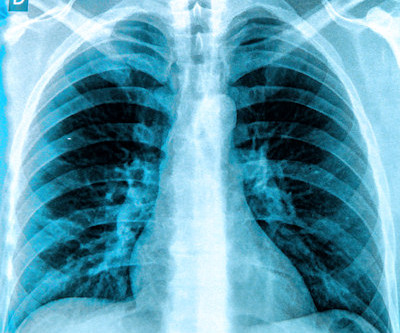
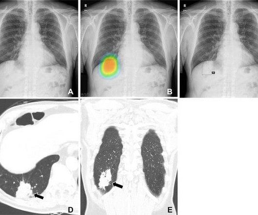
Of the 120 chest radiographs assessed, 60 were from lung cancer patients (32 males) and 60 were controls (36 males). from the Department of Radiology and Institute of Radiation Medicine at Seoul National University College of Medicine in Seoul. “We Patients had a median age of 67 years.
Frontal abdomen radiograph demonstrates foreign body consistent with capsule endoscopy device (pill cam) in descending colon. Approximately 2% of all capsule endoscopies result in CR [ 2 ] The clinical indication for capsule endoscopy is correlated with different rates of CR. Xray of the Week Figure 1. Gastrointest Endosc.
The following panelists of experts share their insights and experiences in the use of WBCT in clinical practice as well as the future direction of the modality. How do you use it in surgical decision making for Hallux Valgus, PCFD, and Trauma (such as Lisfranc and Syndesmosis injuries)?
Assessment of Claimant, Clinical, and Financial Characteristics of Teleradiology Medical Malpractice Cases. Commercially Available Chest Radiograph AI Tools for Detecting Airspace Disease, Pneumothorax, and Pleural Effusion. Effects of low-dose ionizing radiation on genomic instability in interventional radiology workers.
The data will be available through the OSIC Data Repository to AI experts and other collaborators to design algorithms that could potentially identify novel biomarkers and relate radiograph quantifications to clinical indicators, and to disease risk and prognostication factors. Stop and think for a moment about this possibility.
X-ray Also called a radiograph, an X-ray uses radiation to create images of the body. Like the X-ray, a CT scan sends radiation through your body with a much detailed view of its structures. Unlike X-rays, MRIs don’t have the harmful effects of radiation. Dr. Jorge L.
Photoprint from radiograph by W.K. 3) The British Röntgen Society (the first radiology society) was founded in 1897, and many further studies on X-ray usage and the effects of radiation were performed over the following years. (3) It has become one of the most important topics of research and development in clinical radiology. (17)
As we have seen from the past 30 years, imaging has become more powerful, with resolutions continuing to improve whilst radiation doses have simultaneously dropped, and these advances will continue. They will still play a vital role in advising other doctors which scan would be the most appropriate to answer their clinical query.
Patient demographics, aneurysm characteristics, surgical procedures, and clinical outcomes were analyzed. Of the patients with radiographically proven adverse radiation effects (AREs; 15%), 4 were symptomatic. The patterns of recurrence were classified as local, marginal, and distant based on the range of radiation.
We organize all of the trending information in your field so you don't have to. Join 5,000 users and stay up to date on the latest articles your peers are reading.
You know about us, now we want to get to know you!
Let's personalize your content
Let's get even more personalized
We recognize your account from another site in our network, please click 'Send Email' below to continue with verifying your account and setting a password.


Let's personalize your content