This site uses cookies to improve your experience. To help us insure we adhere to various privacy regulations, please select your country/region of residence. If you do not select a country, we will assume you are from the United States. Select your Cookie Settings or view our Privacy Policy and Terms of Use.
Cookie Settings
Cookies and similar technologies are used on this website for proper function of the website, for tracking performance analytics and for marketing purposes. We and some of our third-party providers may use cookie data for various purposes. Please review the cookie settings below and choose your preference.
Used for the proper function of the website
Used for monitoring website traffic and interactions
Cookie Settings
Cookies and similar technologies are used on this website for proper function of the website, for tracking performance analytics and for marketing purposes. We and some of our third-party providers may use cookie data for various purposes. Please review the cookie settings below and choose your preference.
Strictly Necessary: Used for the proper function of the website
Performance/Analytics: Used for monitoring website traffic and interactions
This year’s trip along the Road to RSNA for digital x-ray features a few familiar mileposts – AI for chest x-ray studies, for instance – but notably also significant research into how technology and new techniques can reduce radiation exposure in patients. DAEC system reduces portable x-ray radiation doses Sunday, December 1 | 1:20 p.m.-1:30
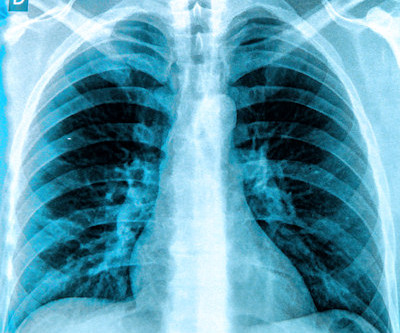
Chest dynamic digital radiography (DDR) may have received a boost toward clinical use in patients with lung disorders, with researchers developing AI to perform time-consuming analysis involved in the technology, according to researchers in New York City.
More than 60% of diagnostic radiology and radiation therapy staff experience workplace violence, according to a study published January 9 in Radiography. Yet no systematic review has been published on the issue in medical radiation science, the authors noted.
12, 2025 Konica Minolta Healthcare Americas, has published a case study by clinicians in the pulmonary and radiology departments at ASST Fatebenefratelli Sacco (Milan, Italy) demonstrating the use of Dynamic Digital Radiography (DDR) to help definitively diagnose diaphragm dysfunction. tim.hodson Fri, 02/14/2025 - 15:14 Feb.12,
Staff operating ionizing radiation equipment in Michigan are now required to meet certain qualifications for active status and employment. We worked with the state to update the language that existed on equipment operation, education, clinical requirements, and credentialing," Aragon explained in the ARRT update.
The company will showcase the clinical analysis of Canon’s Intelligent Noise Reduction (Intelligent NR) that provides superior image quality while lowering radiation dosing in pediatric digital radiography at the Radiological Society of North America Annual Meeting 2023 (RSNA) , McCormick Place Convention Center, Chicago, IL Nov.
Most Influential Radiology Researcher Erik Middlebrooks, MD, Mayo Clinic, Jacksonville, FL Erik Middlebrooks, MD. One of this year's nominations for Most Influential Radiology Researcher is Erik Middlebrooks, MD, of the Mayo Clinic in Jacksonville, FL.
S4-SSPH02-3 | Room S404 In this session on physics in radiography, a study suggests portable x-ray systems outfitted with digital autoexposure control (DAEC) systems enhance image quality and reduce radiation doses. Sunday, December 1 | 1:20 p.m.-1:30 1:30 p.m. | manual vs. 13.4,
Louis asked, “How much ionizing radiation are neonatal patients exposed to during interventional procedures?” S4-SSMK02-2 | Room E353C Generative AI technology shows potential for making surgical planning for total hip arthroplasty (THA) more efficient, according to researchers from the Mayo Clinic. 2:00 p.m. | 3:20 p.m. | 8:30 a.m. |
In an open forum, Yi Xiang Tay, of Singapore University Hospital's radiography and diagnostic imaging department, shared his team's research. In addition, clinical decision support systems were the most evaluated mode of intervention, either integrated or standalone. For more coverage from ECR 2024, please visit our RADCast.
An AI algorithm may make dynamic digital radiography (DDR) more efficient by automatically measuring kinematics involved in certain shoulder injuries, according to a presentation delivered November 29 at RSNA. These images are then processed to visualize joints in motion.
“So we were thinking and asking ourselves, ‘can nonradiologists benefit from AI and chest radiography analysis in this emergency unit set.’ ” Per year, LMU receives between 5,000 and 6,000 orders for chest radiographs for primary diagnosis from the emergency unit alone.
Israel-based Nano-X Imaging (Nanox) will conduct a clinical study evaluating its Nanox.ARC 3D imaging system in a clinical outpatient setting at Clalit Health Services-owned Beilinson Hospital, part of Rabin Medical Center.
Radiography has exploded into a variety of modalities and specialisms from CT to Ultrasound to MRI; all driven by research and development. I’m also passionate about education, research, and (of course) radiography. However, I’m not entirely non-clinical. What is Radiography research and why is it important?
The ability to lower radiation doses without a loss in image quality also has considerable benefits in neonatal and pediatric imaging where imaging at the lowest possible dose is critical. Vincent Chan is president and general manager of digital radiography at Carestream.
Dynamic chest radiography (DCR) shows potential as a tool to investigate lung health in people with cystic fibrosis (CF), according to research published February 13 in Clinical Radiology. and colleagues. Yet FEV1 may not always reflect the severity of the airway obstruction, they noted.
Introduction : In the past decade, veterinary medicine has witnessed a transformative shift with the adoption of digital radiography systems in place of traditional film-based methods. For veterinary professionals contemplating the switch to digital radiography, this article offers essential guidance to ensure a seamless transition.
The following panelists of experts share their insights and experiences in the use of WBCT in clinical practice as well as the future direction of the modality. Cone beam CT, be it weight bearing or not, is equivalent to 3D radiography and that is where most of the healthcare benefits are for patients.
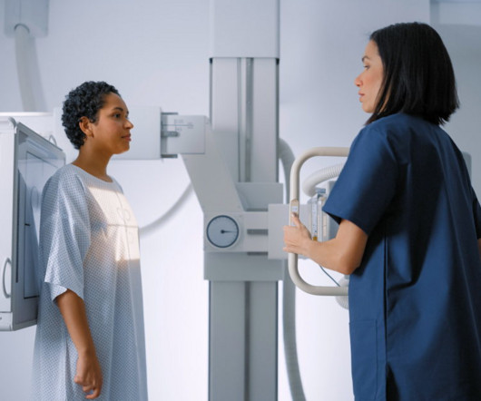
Reading Time: 9 minutes read Carestream solutions help improve patient safety and clinical outcomes. A fundamental goal of radiographers is to complete an imaging exam that provides sufficient information for an accurate clinical diagnosis–and at the lowest possible dose.
announced today several new solutions in digital radiography (DR) and ultrasound that will be introduced at the 2023 Radiology Society of North America (RSNA) Scientific Assembly and Annual Meeting from November 26-30 in Chicago, IL. This capability simplifies clinical workflow and reduces patient exposure to radiation dose.
Shamie Kumar describes how AI fits into a radiology clinical workflow and her perspective on how a clinical radiographer could use this to learn from and enhance their skills. Knowledge, perceptions, and expectations of Artificial intelligence in radiography practice: A global radiography workforce survey. Coakley, Y.
Chest radiography is a common diagnostic tool, but significant training and experience is required to interpret exams correctly,” said lead researcher Louis L. While AI tools are increasingly being approved for use in radiological departments, there is an unmet need to further test them in real-life clinical scenarios,” Dr. Plesner said. “AI
Chapter 2: The Art and Science of Radiography A closer look at the development of radiography, the first X-ray imaging method. How radiography has played a pivotal role in diagnosing bone fractures, identifying foreign bodies, and shaping early healthcare practices.
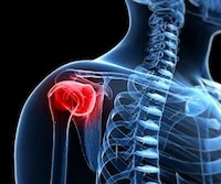
Not only are children more radiosensitive than adults (the cancer risk per unit dose of ionizing radiation is higher), but children also have a longer expected lifetime, which puts them at greater risk of cancer following radiation exposure.(1) The balance of dose and image quality is even more important in pediatric medical imaging.
Radiation oncology clinics face numerous challenges in the present environment, including the simultaneous management of multiple tasks (many of which are manual in nature), various degrees of standardisation, and the potential for errors to impact patient treatment. Healthcare Quarterly. April 2012. doi:10.12927/hcq.2012.22845
Open MRI Equals Greater Comfort, Better Compliance The clinical team at Massac Memorial says the hospital’s Oasis Velocity Open MRI system is boosting efficiency for technologists and offering big benefits to patients. And having that in your own backyard means you’re getting the care you need without having to travel.”
The historical backdrop of Wilhelm Roentgen’s serendipitous discovery and the birth of radiography. Chapter 3: Types of X-ray Technology: Beyond Radiography An exploration of the various modalities and applications of X-ray technology, from radiography to fluoroscopy and computed tomography (CT).
a Sunnyvale, CA-based developer of a next generation artificial intelligence (AI)-based tomosynthesis X-ray imaging system, has reported that its team, which began clinical trials in late 2023, is very pleased with the early ARC60 imaging results, both in terms of depiction of details and consistency of imaging quality.
Chapter 3: The Radiologic Toolbox – Types of X-ray Imaging An exploration of the various types of X-ray imaging, including radiography, fluoroscopy, computed tomography (CT), and more. How each modality serves unique clinical purposes and applications. Real-life case studies illustrating the diagnostic power of X-rays.
The principles of radiation and how X-rays interact with the human body to create diagnostic images. Chapter 3: Types of X-ray Imaging: Beyond Radiography An exploration of the various types of X-ray imaging, including radiography, fluoroscopy, and computed tomography (CT).
Film-Based Radiography: Discuss the era of film-based radiography, highlighting its advantages and limitations. The Dawn of Digital Imaging: Explore the transition from film to digital radiology, emphasizing the benefits of digital technology, including immediate results and reduced radiation exposure.
In this article, we will explore the latest innovations that are shaping the future of dental X-ray imaging, revolutionizing patient care and clinical practices. Digital Radiography: Traditional film-based X-ray imaging is making way for digital radiography.
These lightweight and easily transportable units empower veterinarians to conduct imaging procedures right in the comfort of the animal’s familiar surroundings, whether that be in a clinic, a stable, or even on a farm.
By Professor Louise Rainford, Associate Dean of Radiography, UCD Dublin. And are clinical staff treated the same across disciplines when they make mistakes? Incorrect markers can result in a patient having to undergo a second exam and additional exposure to radiation. Some safety incidents have nothing to do with radiation.
The ability to lower radiation doses without a loss in image quality also has considerable benefits in neonatal and pediatric imaging where imaging at the lowest possible dose is critical. About the Author Vincent Chan is President and General Manager of Digital Radiography at Carestream.
The following panelists of experts share their insights and experiences in the use of WBCT in clinical practice as well as the future direction of the modality. Trauma : In these injuries one would normally go for a standard radiography followed by second-line imaging such as CT.
Arm position is a significant factor influencing radiation exposure to patients during whole-body PET/CT scans, according to a team of radiologic technologists in Japan. More accurate understanding of the degree of difference in radiation exposure dose due to arm position is useful information for dose optimization,” the group wrote.
Assessment of Claimant, Clinical, and Financial Characteristics of Teleradiology Medical Malpractice Cases. Effects of low-dose ionizing radiation on genomic instability in interventional radiology workers. Neuroimaging and Clinical Findings in Healthy Middle-Aged Adults With Mild Traumatic Brain Injury in the PREVENT Dementia Study.
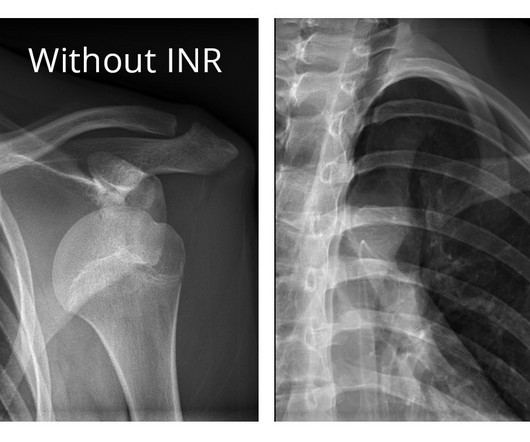
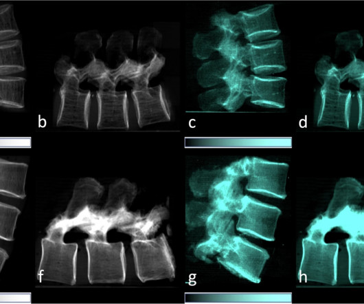
This finding implies the potential application of dark-field imaging to draw conclusions on bone microstructure for predicting bone stability at a lower radiation exposure than in tomographic modalities,” wrote Jon Rischewski, a doctoral candidate at Ludwig Maximilian University of Munich, and colleagues.
Each boasts near perfect sensitivity and negative predictive values for clinically significant acute intracranial processes. Only New Orleans included clinical intoxication, while NEXUS was the only rule to include coagulopathy. versus 99.1%) compared with the original study for clinically significant intracranial injury.
We organize all of the trending information in your field so you don't have to. Join 5,000 users and stay up to date on the latest articles your peers are reading.
You know about us, now we want to get to know you!
Let's personalize your content
Let's get even more personalized
We recognize your account from another site in our network, please click 'Send Email' below to continue with verifying your account and setting a password.


Let's personalize your content