This site uses cookies to improve your experience. To help us insure we adhere to various privacy regulations, please select your country/region of residence. If you do not select a country, we will assume you are from the United States. Select your Cookie Settings or view our Privacy Policy and Terms of Use.
Cookie Settings
Cookies and similar technologies are used on this website for proper function of the website, for tracking performance analytics and for marketing purposes. We and some of our third-party providers may use cookie data for various purposes. Please review the cookie settings below and choose your preference.
Used for the proper function of the website
Used for monitoring website traffic and interactions
Cookie Settings
Cookies and similar technologies are used on this website for proper function of the website, for tracking performance analytics and for marketing purposes. We and some of our third-party providers may use cookie data for various purposes. Please review the cookie settings below and choose your preference.
Strictly Necessary: Used for the proper function of the website
Performance/Analytics: Used for monitoring website traffic and interactions
Intelligent virtual and AI-based collimation features appear to save radiographers time during x-ray image acquisitions – a key function for enabling more patient-focused workflows, according to a recent study. To that end, the researchers conducted an observational study at five clinical sites in Europe and the U.S.
million chest radiographs. The team reported that the algorithm could successfully triage pairs of chest radiographs showing no change while detecting urgent interval changes during longitudinal follow-up. Julianna Czum, MD, from Johns Hopkins University wrote an editorial accompanying the study.
The results of our study demonstrated that preliminary reports created by a multimodal generative AI model could aid radiologists in chest radiograph interpretation in terms of reading time, report quality, and accuracy, noted lead author Eun Kyoung Hong, MD, of Mass General Brigham in Boston.
AI for thoracic imaging includes using it for reading chest radiographs and low-dose chest CT scans for lung cancer screening and for triaging pulmonary embolism on chest CT scans, the group noted. These LLMs offer opportunities ranging from generating text reports from images to explaining examination results to patients."
Some of my radiological heroes would report a staggering 30,000 to 40,000 radiographs a year. Some even [startled gasp] gave up reporting plain radiographs. And they did this alarming juggling act for hour after hour, day after day, demolishing huge piles of film packets. I still don’t know how they did it.
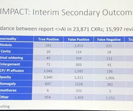
. | S1-SSCH01-5 | E451A This scientific paper may increase overall confidence in the potential of using multimodal AI for tuberculosis (TB) detection, and potentially autonomous reporting, on chest radiographs in certain clinical settings. Stop by the session with your questions.
These included chest radiographs that displayed abnormalities of no clinical significance, which are typically treated as normal. In addition, a thoracic radiologist graded the missed findings by AI and/or the radiology reports as critical, clinically significant, or clinically insignificant.
Chest dynamic digital radiography (DDR) may have received a boost toward clinical use in patients with lung disorders, with researchers developing AI to perform time-consuming analysis involved in the technology, according to researchers in New York City. a) Raw example of a dynamic digital radiograph. (b)
To address this gap in evidence, the researchers developed a decision analytic model to estimate clinical outcomes associated with PSMA-PET versus conventional imaging strategies, namely CT and bone scans.
Moreover, while manual measurement of femoral and tibial length on radiographs has been shown to be reliable, it is a tedious and time-consuming task, the authors added. The study data included 1,874 examinations from 523 pediatric patients aged 0 to 21 who underwent at least two slot-scanning radiographs in routine clinical care.
Middlebrooks, MD, Mayo Clinic, Jacksonville, FL Erik H. Middlebrooks, MD, of the Mayo Clinic in Jacksonville, FL. Given the extensive array of sequences and techniques employed in clinical imaging, we must address the inherent challenges associated with each," he noted. I'm a radiographer,' " Stewart recalled.
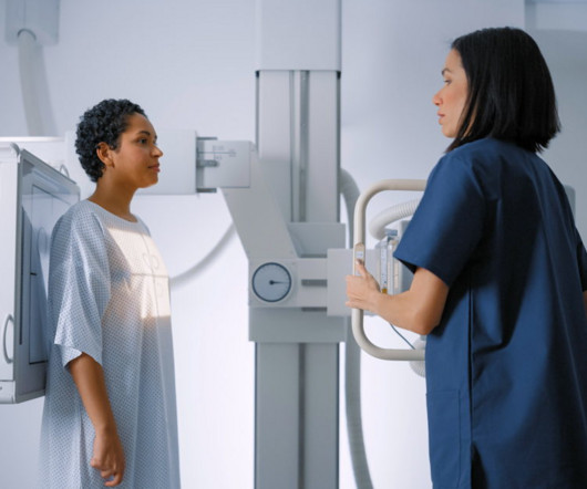
At RSNA 2023, look for AI-driven systems that radiographers can use to help make patient positioning faster and more precise, and bring consistency to the process, all of which help improve image quality and reduce the need for retakes. Even the most skilled radiographers can fail to get positioning just right.
ChatGPT-4 outperformed human clinicians in determining pretest and post-test disease probability after a negative test result involving chest radiographs and mammograms, according to a research letter published December 11 in JAMA Network Open.
. | W3-SSMK08-4 | Room E450A A deep learning-based framework for automated screening of osteoporosis on lumbar spine plain radiographs shows potential as another way to opportunistically make use of imaging studies performed for other indications, according to this presentation.
The deep learning (DL) model may guide clinical decision-making in the management of patients with CAP by identifying high-risk patients who warrant hospitalization and intensive treatment,” concluded first author Eui Jin Hwang, MD, PhD, from the department of radiology at Seoul National University College of Medicine in Korea. Hwang et al.
RSNA attendees focused on practical AI matters will likely appreciate the RSNA's Imaging AI in Practice interoperability demonstration, which will demonstrate at the AI Showcase how the technology can be integrated into radiology workflow in real-world clinical scenarios. 9:40 a.m. | 9:40 a.m. | 10:00 a.m. | 2:00 p.m. | 1:50 p.m. |
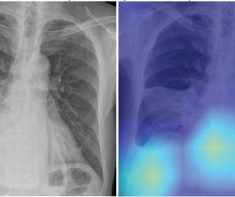
Our findings suggest that artificial intelligence assistance in chest radiograph interpretation may enhance sensitivity without affecting specificity for all readers, regardless of their level of expertise or seniority or the type of abnormality,” wrote first author Souhail Bennani, MD, and colleagues. Image courtesy of Radiology.
Its impact on radiographer workflow ranges from detecting poor image quality on x-ray; automating CT imaging protocols; and for MRI, streamlining workflows for faster scan times, image reconstruction, and using synthetic MRI sequences.
The finding validates fully automated software for use in procedures and advances DDR a step toward clinical use, the technology’s U.S. Representative chest radiographs and graphs of lung signal intensity obtained using DDR for the severe COPD group. and Japanese developers wrote. The subject is a 77-year-old male with VC of 1.68
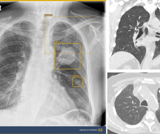
Clinical centers rarely have the necessary resources and personnel to evaluate and compare multiple products prior to purchase,” the group wrote. A) Radiograph in a man (age, 72 years) with a nodule present (reference standard score, 100) shows a true-positive result based on the average algorithm scores. (B)
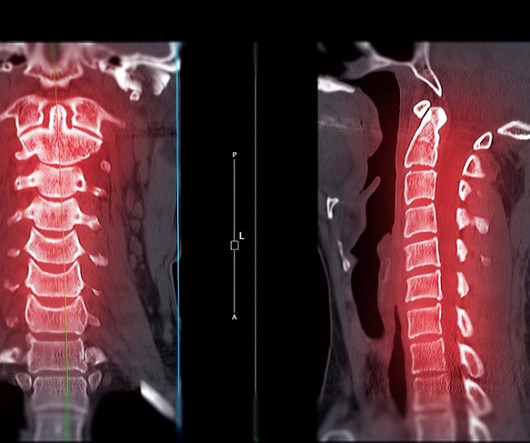
A highly accurate prediction rule for cervical spine injuries in children reduces the use of CT scans by over 50% without missing clinically significant injuries or increasing the use of unnecessary X-rays.
The rules also establish initial and continuing education requirements for limited-scope radiographers and radiologist assistants. We worked with the state to update the language that existed on equipment operation, education, clinical requirements, and credentialing," Aragon explained in the ARRT update.
Deep-learning models could have potential as predictive tools for breast cancer prognosis, a study published January 17 in Clinical Breast Cancer has found. From there, the team used clinical data and imaging characteristics to select independent prognostic factors to establish a clinical model.
The overhead tube crane system is designed to deliver consistent, efficient and comfortable patient exams, enhance technologist’s workflow, deliver clinical confidence, and help to keep imaging departments running smoothly. launch of its latest addition to its advanced digital radiography suites, the D-EVO Suite OTCx.
Kevin Daynes, MD, filed suit with the New South Wales Supreme Court after he was fired in May 2022 from the I-MED Radiology Cairns clinic for "bullying conduct and victimization" complaints from a female radiographer and a sexual harassment complaint from a female technologist, the outlet reported. million Australian dollars ($2.9
Most Influential Radiology Researcher Erik Middlebrooks, MD, Mayo Clinic, Jacksonville, FL Erik Middlebrooks, MD. One of this year's nominations for Most Influential Radiology Researcher is Erik Middlebrooks, MD, of the Mayo Clinic in Jacksonville, FL.
The study, published in the journal Diagnostics, highlights the clinical value of DDR through its unique ability to evaluate diaphragm movement in real time and integrate dynamic functional information with static anatomical data to provide a quantitative assessment of diaphragmatic movement, including excursion and speed.
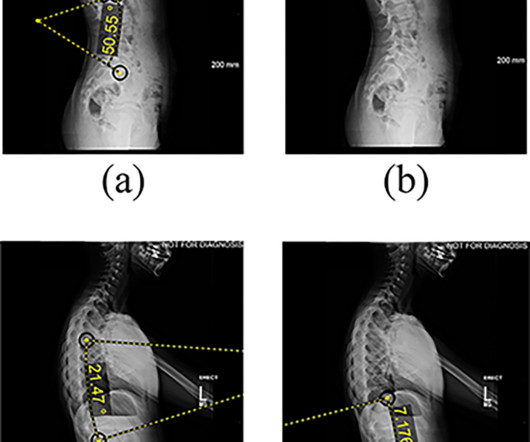
The current model has potential to serve as a valuable clinical tool, providing insight into the optimal timing of intervention and surgical planning parameters,” the group wrote. Features extracted from lateral radiographs. (a) AVBT was approved in the U.S. The full study can be found here.
Shamie Kumar describes how AI fits into a radiology clinical workflow and her perspective on how a clinicalradiographer could use this to learn from and enhance their skills. If the AI findings are seen in PACS, how many radiographers actually log into PACS after taking a scan or X-ray? Can Radiographers Up-Skill?
This competition demonstrated the value of AI in detecting and localizing many pathologies in chest radiographs by simulating the real work situations of radiologists,” the group wrote. The study was published February 8 in the Journal of Imaging Informatics in Medicine. The full study is available here.
This is a potentially practice-changing trial,” said Oliver Sartor, MD, a medical oncologist and director of radiopharmaceutical trials at the Mayo Clinic in Rochester, MN. Radiographic progression-free survival (rPFS) was the primary endpoint of the study, while overall survival was the key secondary endpoint.
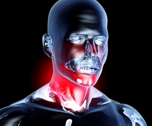
Radiologists can help reduce unnecessary follow-up work in patients with suspected oropharynx cancer by analyzing certain metrics on PET/CT scans, according to a team of head and neck surgeons at the Mayo Clinic in Rochester, MN.
Initial results from currently ongoing clinical studies suggest that dark-field chest x-ray can be useful for detecting and quantification of pulmonary emphysema , and for assessing COVID-19 pneumonia , the authors wrote. The article was published March 7 in IEEE Transactions on Medical Imaging.
DDR is an emerging imaging technique that uses a pulsed x-ray source to acquire a series of radiographs at six to 15 frames per second. These images are then processed to visualize joints in motion.
Communication among practitioners: Effective communication between radiographers, radiologists, and child-life specialists is needed to discuss scan protocols, streamline the exam, and minimize table time. Having support from a child-life specialist inside the scan room can help children stay calm and cooperative.
“So we were thinking and asking ourselves, ‘can nonradiologists benefit from AI and chest radiography analysis in this emergency unit set.’ ” Per year, LMU receives between 5,000 and 6,000 orders for chest radiographs for primary diagnosis from the emergency unit alone.
The technology figured prominently in five Minnies categories, including Hottest Clinical Procedure. Pickhardt believes using additional clinical data on CT imaging could save healthcare costs, especially when AI tools are used in parallel, suggesting it is a cost-effective or even cost-saving strategy for personalized or precision medicine.
Others on the team include lead investigator David Baldwin, MD, chair of NHS England’s Clinical Expert Group for Lung Cancer and a respiratory consultant at Nottingham University Hospitals, cardiothoracic radiologist Indrajeet Das, MD, and Prof. UCLH has produced 9,217 chest x-rays from 8,072 patients.
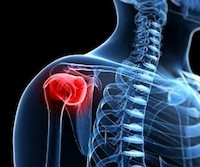
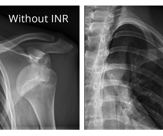
Researchers at the Mayo Clinic in Rochester, MN, have taken a first step in using AI to automatically classify and organize shoulder x-rays on a large scale, according to a recent study. To date, most registries consist primarily of clinical data, and either completely lack or contain very limited medical imaging information.
The company will showcase the clinical analysis of Canon’s Intelligent Noise Reduction (Intelligent NR) that provides superior image quality while lowering radiation dosing in pediatric digital radiography at the Radiological Society of North America Annual Meeting 2023 (RSNA) , McCormick Place Convention Center, Chicago, IL Nov.
Tay said while radiology professional societies have advocated incorporating evidence-based imaging referral guidelines into clinical practice, the extent of progress in implementing these guidelines varies worldwide. In addition, clinical decision support systems were the most evaluated mode of intervention, either integrated or standalone.
The following panelists of experts share their insights and experiences in the use of WBCT in clinical practice as well as the future direction of the modality. Whenever bilateral standing radiographs would have been needed, a WBCT was performed instead. How Frequently Do You Use It In Practice?
Assessment of Claimant, Clinical, and Financial Characteristics of Teleradiology Medical Malpractice Cases. Commercially Available Chest Radiograph AI Tools for Detecting Airspace Disease, Pneumothorax, and Pleural Effusion. Generative Artificial Intelligence for Chest Radiograph Interpretation in the Emergency Department.
Together with our radiographers, I learned to scan cardiac patients and learned special anatomy from pediatric cardiologists and pediatric cardiac surgeons." At the time, there was little training for, say, cardiac MRI. But he persevered. Imaging was so important [for cardiac indications], that I decided to become a radiologist," he said.
We organize all of the trending information in your field so you don't have to. Join 5,000 users and stay up to date on the latest articles your peers are reading.
You know about us, now we want to get to know you!
Let's personalize your content
Let's get even more personalized
We recognize your account from another site in our network, please click 'Send Email' below to continue with verifying your account and setting a password.


Let's personalize your content