This site uses cookies to improve your experience. To help us insure we adhere to various privacy regulations, please select your country/region of residence. If you do not select a country, we will assume you are from the United States. Select your Cookie Settings or view our Privacy Policy and Terms of Use.
Cookie Settings
Cookies and similar technologies are used on this website for proper function of the website, for tracking performance analytics and for marketing purposes. We and some of our third-party providers may use cookie data for various purposes. Please review the cookie settings below and choose your preference.
Used for the proper function of the website
Used for monitoring website traffic and interactions
Cookie Settings
Cookies and similar technologies are used on this website for proper function of the website, for tracking performance analytics and for marketing purposes. We and some of our third-party providers may use cookie data for various purposes. Please review the cookie settings below and choose your preference.
Strictly Necessary: Used for the proper function of the website
Performance/Analytics: Used for monitoring website traffic and interactions
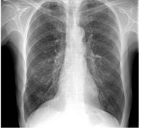
A group in South Korea has validated a generative AI model that could reduce reading times and increase chest x-ray reporting accuracy, according to a study published March 11 in Radiology. Five radiologists interpreted the chest radiographs in two sessions: without AI-generated reports and with AI-generated reports as preliminary reports.
Intelligent virtual and AI-based collimation features appear to save radiographers time during x-ray image acquisitions – a key function for enabling more patient-focused workflows, according to a recent study. To that end, the researchers conducted an observational study at five clinical sites in Europe and the U.S.
The use of AI in thoracic imaging has begun to demonstrate "cumulative evidence of effectiveness," but more testing and research are needed to determine its feasibility for this application, according to a commentary published February 25 in Radiology. van Beek, MD, of the University of Edinburgh in the U.K. in an accompanying editorial.
Some of my radiological heroes would report a staggering 30,000 to 40,000 radiographs a year. I remember the crossing of this particular radiological Rubicon about 15 years ago here in the U.K. Some even [startled gasp] gave up reporting plain radiographs. I still don’t know how they did it. So what happened?
With 100% of precincts now reporting, we’re finally ready to declare this year’s winners in our annual awards program recognizing excellence in radiology. The voters also zeroed in on the ongoing shortage of radiologists as the Biggest Threat to Radiology. Most Influential Radiology Researcher Minnies 2024 Winner: Erik H.
Charlene Liew, MD, director of cardiothoracic imaging at Changi General Hospital in Singapore, outlined the current state of AI in radiology and where it is going in a presentation called "AI in Radiology: The Past Informs the Future." Food and Drug Administration (FDA) were for radiology indications.
Each year, the Minnies award winners reflect the current challenges, issues, and advances in radiology. The technology figured prominently in five Minnies categories, including Hottest Clinical Procedure. And for the first time since 2018, physician burnout did not win the Minnies award for Biggest Threat to Radiology.
A commercially available AI algorithm shows potential for off-label use as a way to generate automatic reports for “unremarkable” chest x-rays, according to a study published August 20 in Radiology. These included chest radiographs that displayed abnormalities of no clinical significance, which are typically treated as normal.
Who made it to the final round in the 2024 edition of the Minnies, AuntMinnie.com 's event recognizing excellence in radiology? Most Influential Radiology Researcher Erik Middlebrooks, MD, Mayo Clinic, Jacksonville, FL Erik Middlebrooks, MD. The Minnies finalists were drawn from over 200 candidates across 15 categories.
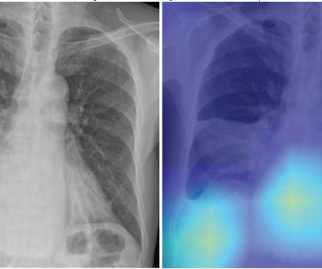
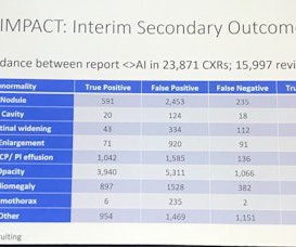
AI assistance can improve the detection accuracy of thoracic abnormalities on chest x-rays across radiologists with varying levels of expertise, according to a study published December 12 in Radiology. Image courtesy of Radiology. In a retrospective study, a commercially available algorithm (ChestView, v.
An AI model has delivered a long overdue update of pediatric bone growth predictions used in x-ray imaging to monitor scoliosis, according to a study published April 30 in Radiology. The lower extremity measurement pipeline is illustrated using a representative slot-scanning radiograph. Image courtesy of Radiology.
The new rules now in effect in Michigan help ensure that only medical radiologic technologists with the appropriate education and training can operate medical imaging equipment, according to an American Registry of Radiologic Technologists (ARRT) legislative update.
AI’s capability and potential to enhance the practice of radiology will take center stage at RSNA 2023. AI highlights clinically significant prostate cancer on MRI Sunday, November 26 | 9:30 a.m.-9:40 AI highlights clinically significant prostate cancer on MRI Sunday, November 26 | 9:30 a.m.-9:40 9:40 a.m. | 9:40 a.m. |
This year, hundreds of candidates have been selected as semifinalists for 14 categories, ranging from Most Influential Radiology Researcher to Best New Radiology Software. The semifinalist list was compiled based on nominations from members of AuntMinnie.com. Winners will be selected by our expert panel in two rounds of voting.
The finding validates fully automated software for use in procedures and advances DDR a step toward clinical use, the technology’s U.S. The study was published in the December 2024 issue of the European Journal of Radiology Open. Image courtesy of the European Journal of Radiology Open. and Japanese developers wrote.
At RSNA 2023, look for AI-driven systems that radiographers can use to help make patient positioning faster and more precise, and bring consistency to the process, all of which help improve image quality and reduce the need for retakes. Even the most skilled radiographers can fail to get positioning just right.
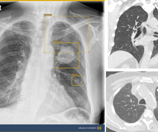
Four out of seven commercially available AI algorithms for detecting lung nodules on x-rays performed better than human readers, while two algorithms for predicting bone age fell short, in a study published January 9 in Radiology. Their performance was compared to reads by 17 radiologists and radiology residents with varying experience.
Radiology plays a crucial role in modern medicine, answering essential diagnostic questions pertaining to nearly every pathology facing clinicians. The Impact on Patients and Clinical Management A 2024 study in the Journal of Clinical Imaging found that 68% of surveyed radiology practices had unreported radiology exams.
Implementing imaging referral guidelines not only supports value-based radiology but makes it easier to communicate with patients about low-value services, according to findings presented February 29 at ECR 2024. It was the endnote of a series of sessions focused on optimizing radiology services.
In this AJR accepted manuscript , a DL model was developed in 7,105 patients via one institution from March 2013 to December 2019 (3:1:1 allocation to training, validation, and internal test sets) to predict risk of all-cause mortality within 30 days after CAP diagnosis using patients’ initial chest radiograph. CURB-65 score). “The
Presenting the research on November 28 at RSNA 2023, Jan Rudolph, MD, from the department of radiology at University Hospital LMU Munich said nonradiologists can significantly benefit from AI assistance in emergency-related chest x-ray analysis. “If Nonradiology residents 0.78 Nonradiology residents 0.78 Nonradiology residents 0.78
Kevin Daynes, MD, filed suit with the New South Wales Supreme Court after he was fired in May 2022 from the I-MED Radiology Cairns clinic for "bullying conduct and victimization" complaints from a female radiographer and a sexual harassment complaint from a female technologist, the outlet reported. million U.S.). million U.S.).
Radiologic technologist educators need AI training and guidance, time to develop educational resources, and support from higher education institutions to improve on the teaching and use of AI, according to a recent study. In a survey of all U.S. The study was published July 13 in the Journal of Medical Imaging and Radiation Sciences.
Radiologists can help reduce unnecessary follow-up work in patients with suspected oropharynx cancer by analyzing certain metrics on PET/CT scans, according to a team of head and neck surgeons at the Mayo Clinic in Rochester, MN.
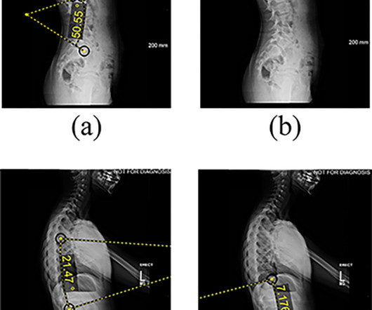
The current model has potential to serve as a valuable clinical tool, providing insight into the optimal timing of intervention and surgical planning parameters,” the group wrote. Features extracted from lateral radiographs. (a) AVBT was approved in the U.S. The full study can be found here.
12, 2025 Konica Minolta Healthcare Americas, has published a case study by clinicians in the pulmonary and radiology departments at ASST Fatebenefratelli Sacco (Milan, Italy) demonstrating the use of Dynamic Digital Radiography (DDR) to help definitively diagnose diaphragm dysfunction. tim.hodson Fri, 02/14/2025 - 15:14 Feb.12,
A range of federal and state legislation and other developments in the works could have a significant impact on radiologic technologists and registered radiology assistants, either supporting and strengthening these professions or weakening them. Existing law, the Radiologic Technology Act, prohibits certain activities.
When we looked at the CTs which were done based on the radiology report saying this is suspicious of cancer, please fast-track it, there was actually quite a difference," Woznitza said. UCLH has produced 9,217 chest x-rays from 8,072 patients. Scans undergo data cleaning, Woznitza explained to retain only scans necessary for compliance.
Initial results from currently ongoing clinical studies suggest that dark-field chest x-ray can be useful for detecting and quantification of pulmonary emphysema , and for assessing COVID-19 pneumonia , the authors wrote. The article was published March 7 in IEEE Transactions on Medical Imaging.
The company will showcase the clinical analysis of Canon’s Intelligent Noise Reduction (Intelligent NR) that provides superior image quality while lowering radiation dosing in pediatric digital radiography at the Radiological Society of North America Annual Meeting 2023 (RSNA) , McCormick Place Convention Center, Chicago, IL Nov.
This competition demonstrated the value of AI in detecting and localizing many pathologies in chest radiographs by simulating the real work situations of radiologists,” the group wrote. The study was published February 8 in the Journal of Imaging Informatics in Medicine. The full study is available here.
Together with our radiographers, I learned to scan cardiac patients and learned special anatomy from pediatric cardiologists and pediatric cardiac surgeons." Acknowledge that cardiac imaging is … not only about radiology," he said. At the time, there was little training for, say, cardiac MRI. But he persevered.
Shamie Kumar describes how AI fits into a radiologyclinical workflow and her perspective on how a clinicalradiographer could use this to learn from and enhance their skills. If the AI findings are seen in PACS, how many radiographers actually log into PACS after taking a scan or X-ray? Can Radiographers Up-Skill?
Reading Time: 9 minutes read Carestream solutions help improve patient safety and clinical outcomes. A fundamental goal of radiographers is to complete an imaging exam that provides sufficient information for an accurate clinical diagnosis–and at the lowest possible dose.
Communication among practitioners: Effective communication between radiographers, radiologists, and child-life specialists is needed to discuss scan protocols, streamline the exam, and minimize table time. Having support from a child-life specialist inside the scan room can help children stay calm and cooperative.
AI algorithms appear to have clinical value based on detecting normal x-rays – that is, by flagging chest x-rays as normal versus abnormal, they may reduce reading times for radiologists, according to research presented recently at the RSNA meeting in Chicago. In a session on chest imaging, scientists from AI developers Lunit and DeepTek.ai
Harrisons technology in radiology and pathology assists clinicians with identifying signs of cancer and other critical illnesses earlier, improving treatment decisions and patient outcomes. meets this need by developing clinical-grade AI models designed to improve capacity. Averaged across all findings on chest radiographs.) [2]AIDE
milla1cf Mon, 06/24/2024 - 20:34 June 24, 2024 — The Radiological Society of North America ( RSNA ) announced the 2023 impact factors today for its suite of journals. Radiology, RSNA’s leading medical imaging research journal, was cited nearly 60,000 times, according to the Clarivate Analytics Journal Citation Reports.
MRI helps clinicians assess the neural involvement in endometriosis and could help them prevent irreversible nerve damage and chronic sensorimotor neuropathy in women suffering from the condition, Cleveland Clinic researchers have reported. The team's review of MRI's role for this indication was published January 3 in RadioGraphics.
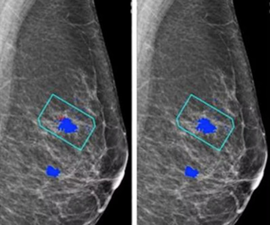
For the AI study, two views of each breast (four images) are taken to be evaluated independently either by the AI system plus a single reader or by a radiologist/radiographer as a first reader and second reader using the two-reader model. Further, radiology researchers at the University of Aberdeen in the U.K.
In addition, an in-person demonstration called Radiology Reimagined: AI, innovation, and interoperability in practice is designed to showcase new technologies and communications standards needed to integrate AI into the diagnostic workflow, according to the RSNA. . | 10:50 a.m. | 1:50 p.m. | 8:30 a.m. | 9:40 a.m. | 1:50 p.m. |
When the radiology department at Cork University Hospital in the south coast of Ireland took the lead in developing an autism-friendly patient experience, they observed that it took less time to complete imaging studies for children with autism, according to a February 28 session at ECR 2024. Implementing sign language (in this case, Lamh).
BoneView is a clinical decision support AI-based software solution designed to efficiently identify bone fractures by highlighting areas of interest on an x-ray image. Food and Drug Administration (FDA)-cleared computer-assisted detection tool (CADe) was trained on tens of thousands of radiographic images.
MyImageDx provides the best of both worlds in terms of radiology education. MyImageDx providers radiology educators access to a personalized imaging educational platform without having to build it in-house.
We organize all of the trending information in your field so you don't have to. Join 5,000 users and stay up to date on the latest articles your peers are reading.
You know about us, now we want to get to know you!
Let's personalize your content
Let's get even more personalized
We recognize your account from another site in our network, please click 'Send Email' below to continue with verifying your account and setting a password.


Let's personalize your content