This site uses cookies to improve your experience. To help us insure we adhere to various privacy regulations, please select your country/region of residence. If you do not select a country, we will assume you are from the United States. Select your Cookie Settings or view our Privacy Policy and Terms of Use.
Cookie Settings
Cookies and similar technologies are used on this website for proper function of the website, for tracking performance analytics and for marketing purposes. We and some of our third-party providers may use cookie data for various purposes. Please review the cookie settings below and choose your preference.
Used for the proper function of the website
Used for monitoring website traffic and interactions
Cookie Settings
Cookies and similar technologies are used on this website for proper function of the website, for tracking performance analytics and for marketing purposes. We and some of our third-party providers may use cookie data for various purposes. Please review the cookie settings below and choose your preference.
Strictly Necessary: Used for the proper function of the website
Performance/Analytics: Used for monitoring website traffic and interactions
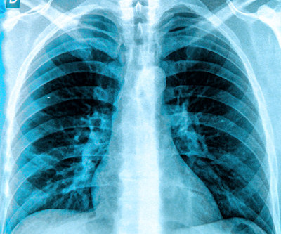
A group in South Korea has validated a generative AI model that could reduce reading times and increase chest x-ray reporting accuracy, according to a study published March 11 in Radiology. Nonetheless, the era of generative AI in radiology holds great promise, they wrote.
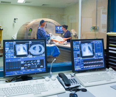
Intelligent virtual and AI-based collimation features appear to save radiographers time during x-ray image acquisitions – a key function for enabling more patient-focused workflows, according to a recent study. To that end, the researchers conducted an observational study at five clinical sites in Europe and the U.S.
A commercially available AI algorithm shows potential for off-label use as a way to generate automatic reports for “unremarkable” chest x-rays, according to a study published August 20 in Radiology. These included chest radiographs that displayed abnormalities of no clinical significance, which are typically treated as normal.
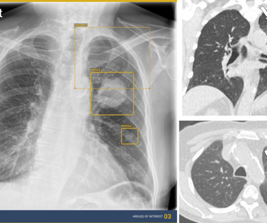
AI assistance can improve the detection accuracy of thoracic abnormalities on chest x-rays across radiologists with varying levels of expertise, according to a study published December 12 in Radiology. About 50% of the x-rays had abnormal findings. AI detected a lung mass, a lung nodule, and a small pneumothorax.
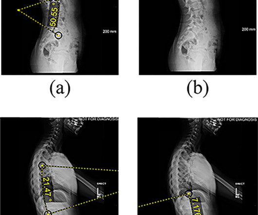
An AI model has delivered a long overdue update of pediatric bone growth predictions used in x-ray imaging to monitor scoliosis, according to a study published April 30 in Radiology. Full-body biplanar slot scanning is a type of low-dose digital x-ray imaging used to monitor scoliosis. Image courtesy of Radiology.
German developers of dark-field chest x-ray appear to have overcome a technical limitation of the technology – namely, adjusting for photon scattering caused by interferometers used in the experimental system. The article was published March 7 in IEEE Transactions on Medical Imaging. Access to the full article is available here.
Nearly 72,000 chest x-rays had been randomized as of November 25 (the study is open through December 31), with the two primary outcomes of the trial being time to diagnosis of lung cancer and time from chest x-ray to CT by prioritizing abnormals. UCLH has produced 9,217 chest x-rays from 8,072 patients.
In a study described as a “competition between radiologists,” participants tasked with identifying abnormal findings on chest x-rays performed better with AI assistance than without AI assistance – though not by much and not in all cases, according to a group in Nanjing, Jiangsu, China. The full study is available here.
Reading Time: 10 minutes read By Henry Williams, Carestream Area Vice President, Sales Western Nowadays, with hospital budgetary restrictions at the forefront of the purchasing decision making process, it seems like the X-Ray market, like everything else, is not immune to the current state of the economy. But is that really the case?
CHICAGO -- German researchers are testing ways to support nonradiologists in interpreting chest x-rays in emergency settings using an AI assistant. Cohort characteristics included pathology prevalence that was suspected pleural effusion, pneumonia, pneumothorax, and lesions. Nonradiology residents 0.78
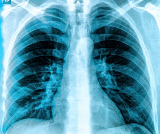
. | S1-SSCH01-5 | E451A This scientific paper may increase overall confidence in the potential of using multimodal AI for tuberculosis (TB) detection, and potentially autonomous reporting, on chest radiographs in certain clinical settings. Stop by the session with your questions.
Chest dynamic digital radiography (DDR) may have received a boost toward clinical use in patients with lung disorders, with researchers developing AI to perform time-consuming analysis involved in the technology, according to researchers in New York City. a) Raw example of a dynamic digital radiograph. (b)
million chest radiographs. The team reported that the algorithm could successfully triage pairs of chest radiographs showing no change while detecting urgent interval changes during longitudinal follow-up. Julianna Czum, MD, from Johns Hopkins University wrote an editorial accompanying the study.
Four out of seven commercially available AI algorithms for detecting lung nodules on x-rays performed better than human readers, while two algorithms for predicting bone age fell short, in a study published January 9 in Radiology. Project AIR is an ongoing cohort study aimed at filling this gap, the authors wrote.
An AI model for x-ray imaging could help clinicians plan treatment other than spinal fusions in patients with adolescent idiopathic scoliosis, according to research published January 14 in PLOS One. For all patients, spinal x-rays were taken at six visits, from patients’ first standing x-ray to their most recent follow-up exam.
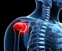
Researchers at the Mayo Clinic in Rochester, MN, have taken a first step in using AI to automatically classify and organize shoulder x-rays on a large scale, according to a recent study. To date, most registries consist primarily of clinical data, and either completely lack or contain very limited medical imaging information.
The finding validates fully automated software for use in procedures and advances DDR a step toward clinical use, the technology’s U.S. DDR is a novel functional imaging technique that uses sequential images obtained by a pulsed x-ray generator and a flat panel detector with a large field of view. and Japanese developers wrote.
DDR is an emerging imaging technique that uses a pulsed x-ray source to acquire a series of radiographs at six to 15 frames per second. These images are then processed to visualize joints in motion.
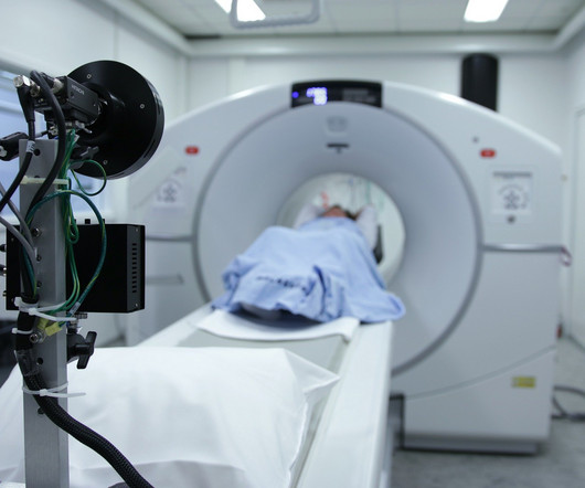
For X-rays, it usually takes less than 10 minutes. This article will talk about the different diagnostic imaging methods such as X-rays, CT scans, Ultrasound, and MRI. X-ray Also called a radiograph, an X-ray uses radiation to create images of the body.
RSNA attendees focused on practical AI matters will likely appreciate the RSNA's Imaging AI in Practice interoperability demonstration, which will demonstrate at the AI Showcase how the technology can be integrated into radiology workflow in real-world clinical scenarios. 9:40 a.m. | 9:40 a.m. | 10:00 a.m. | 2:00 p.m. | 1:50 p.m. |
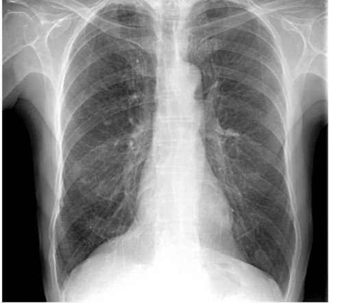
AI algorithms appear to have clinical value based on detecting normal x-rays – that is, by flagging chest x-rays as normal versus abnormal, they may reduce reading times for radiologists, according to research presented recently at the RSNA meeting in Chicago.
Its impact on radiographer workflow ranges from detecting poor image quality on x-ray; automating CT imaging protocols; and for MRI, streamlining workflows for faster scan times, image reconstruction, and using synthetic MRI sequences.
Assessment of Claimant, Clinical, and Financial Characteristics of Teleradiology Medical Malpractice Cases. Commercially Available Chest Radiograph AI Tools for Detecting Airspace Disease, Pneumothorax, and Pleural Effusion. Generative Artificial Intelligence for Chest Radiograph Interpretation in the Emergency Department.
Tay said while radiology professional societies have advocated incorporating evidence-based imaging referral guidelines into clinical practice, the extent of progress in implementing these guidelines varies worldwide. In addition, clinical decision support systems were the most evaluated mode of intervention, either integrated or standalone.
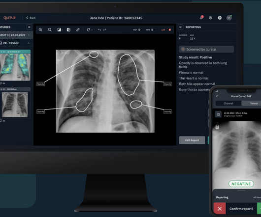
Augmento X-Ray is designed to significantly reduce radiologist workload and improve the quality of chest X-ray reporting. Chest radiography is the most common medical imaging tool used in routine clinical practices to identify different disease findings. billion annual X-rays performed, 1.5
Reading Time: 9 minutes read Mid-cycle refresh can increase clinical, operational, and security benefits. How would you rate the value you are getting from your current X-ray imaging equipment? Does it support new medical imaging software that can help improve clinical outcomes?
Qure’s chest X-ray based qXR-LN uses artificial intelligence to identify and localize lung nodules, marking another significant milestone for the organization, strengthening its standing as a pioneer in the realm of AI-powered advancements for plain film radiography and medical imaging.
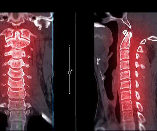
A highly accurate prediction rule for cervical spine injuries in children reduces the use of CT scans by over 50% without missing clinically significant injuries or increasing the use of unnecessary X-rays.
S1-SSCH01-5 | E451A This scientific paper may increase overall confidence in the potential of using multimodal AI for tuberculosis (TB) detection, and potentially autonomous reporting, on chest radiographs in certain clinical settings. Chest x-ray AI performs well in postmarketing surveillance study Tuesday, December 3 | 1:30 p.m.-1:40
The study, published in the journal Diagnostics, highlights the clinical value of DDR through its unique ability to evaluate diaphragm movement in real time and integrate dynamic functional information with static anatomical data to provide a quantitative assessment of diaphragmatic movement, including excursion and speed.
. | W3-SSMK08-4 | Room E450A A deep learning-based framework for automated screening of osteoporosis on lumbar spine plain radiographs shows potential as another way to opportunistically make use of imaging studies performed for other indications, according to this presentation.
At RSNA 2023, look for AI-driven systems that radiographers can use to help make patient positioning faster and more precise, and bring consistency to the process, all of which help improve image quality and reduce the need for retakes. Even the most skilled radiographers can fail to get positioning just right.
R1-SSCH09-5 | Room E352 An AI algorithm can find radiographic markers for osteoporosis that are common but often not reported on radiology reports, according to this scientific paper. The dataset consisted of 519 chest x-rays from patients ages 65 and older, collected from outpatient clinics. 9:00 a.m. |
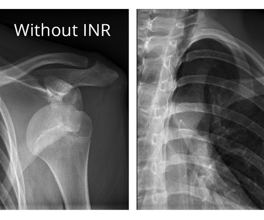
The company will showcase the clinical analysis of Canon’s Intelligent Noise Reduction (Intelligent NR) that provides superior image quality while lowering radiation dosing in pediatric digital radiography at the Radiological Society of North America Annual Meeting 2023 (RSNA) , McCormick Place Convention Center, Chicago, IL Nov.
It uses AI to analyze CT scans, X-rays and pathology slides, supporting clinicians in detecting and diagnosing medical conditions faster and more accurately. meets this need by developing clinical-grade AI models designed to improve capacity. Averaged across all findings on chest radiographs.) [2]AIDE Published 2021.
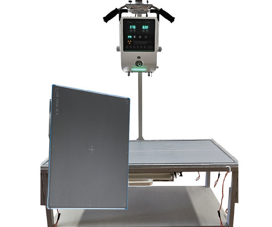
The overhead tube crane system is designed to deliver consistent, efficient and comfortable patient exams, enhance technologist’s workflow, deliver clinical confidence, and help to keep imaging departments running smoothly. X-rays are the most widely used diagnostic tests, accounting for 60% of all imaging studies conducted.
X-ray systems are a fundamental part of this, but the choice between a mobile X-ray system and a stationary one can greatly impact the workflow and efficiency of your clinic. The Elite 125100 Portable X-Ray Machine is an all-in-one portable X-ray machine that includes a 5 kW X-ray power unit.
Reading Time: 9 minutes read Carestream solutions help improve patient safety and clinical outcomes. A fundamental goal of radiographers is to complete an imaging exam that provides sufficient information for an accurate clinical diagnosis–and at the lowest possible dose.
BoneView is a clinical decision support AI-based software solution designed to efficiently identify bone fractures by highlighting areas of interest on an x-ray image. It pinpoints fractures, effusions, dislocations, and bone lesions, and is considered a global bone trauma x-ray interpretation standard, according to Gleamer.
Dynamic chest radiography (DCR) shows potential as a tool to investigate lung health in people with cystic fibrosis (CF), according to research published February 13 in Clinical Radiology. and colleagues. Yet FEV1 may not always reflect the severity of the airway obstruction, they noted.
Too much noise, too much pain, and too much information thrown at them summarize what children with autism can feel in the hospital, explained Martin Peyton, a clinical specialist and radiographer, who presented his hospital's quality initiative. When the child is overstimulated, they are unwilling to assist in the short term.
Detecting CSIs in a clinical setting often requires imaging such as X-rays and computed tomography (CT) scans, both of which expose children to radiation, which can cause other health issues over time.
. | R1-SSCH09-3 | Room E352 This scientific presentation will present external validation results for a deep-learning model in identifying individuals at high risk of incident chronic obstructive pulmonary disease (COPD) on routine outpatient chest x-rays (CXR).
Shamie Kumar describes how AI fits into a radiology clinical workflow and her perspective on how a clinicalradiographer could use this to learn from and enhance their skills. If the AI findings are seen in PACS, how many radiographers actually log into PACS after taking a scan or X-ray?
We organize all of the trending information in your field so you don't have to. Join 5,000 users and stay up to date on the latest articles your peers are reading.
You know about us, now we want to get to know you!
Let's personalize your content
Let's get even more personalized
We recognize your account from another site in our network, please click 'Send Email' below to continue with verifying your account and setting a password.


Let's personalize your content