This site uses cookies to improve your experience. To help us insure we adhere to various privacy regulations, please select your country/region of residence. If you do not select a country, we will assume you are from the United States. Select your Cookie Settings or view our Privacy Policy and Terms of Use.
Cookie Settings
Cookies and similar technologies are used on this website for proper function of the website, for tracking performance analytics and for marketing purposes. We and some of our third-party providers may use cookie data for various purposes. Please review the cookie settings below and choose your preference.
Used for the proper function of the website
Used for monitoring website traffic and interactions
Cookie Settings
Cookies and similar technologies are used on this website for proper function of the website, for tracking performance analytics and for marketing purposes. We and some of our third-party providers may use cookie data for various purposes. Please review the cookie settings below and choose your preference.
Strictly Necessary: Used for the proper function of the website
Performance/Analytics: Used for monitoring website traffic and interactions
Intelligent virtual and AI-based collimation features appear to save radiographers time during x-ray image acquisitions – a key function for enabling more patient-focused workflows, according to a recent study. The study was published online May 18 in Radiography. Finally, VC was used to collimate in 2.4%
12, 2025 Konica Minolta Healthcare Americas, has published a case study by clinicians in the pulmonary and radiology departments at ASST Fatebenefratelli Sacco (Milan, Italy) demonstrating the use of Dynamic Digital Radiography (DDR) to help definitively diagnose diaphragm dysfunction. tim.hodson Fri, 02/14/2025 - 15:14 Feb.12,
Chest dynamic digital radiography (DDR) may have received a boost toward clinical use in patients with lung disorders, with researchers developing AI to perform time-consuming analysis involved in the technology, according to researchers in New York City. a) Raw example of a dynamic digital radiograph. (b)
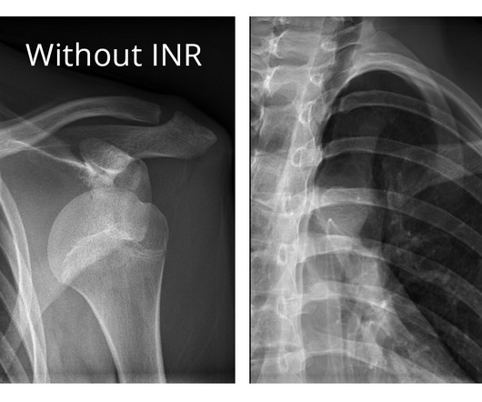
The company will showcase the clinical analysis of Canon’s Intelligent Noise Reduction (Intelligent NR) that provides superior image quality while lowering radiation dosing in pediatric digital radiography at the Radiological Society of North America Annual Meeting 2023 (RSNA) , McCormick Place Convention Center, Chicago, IL Nov.
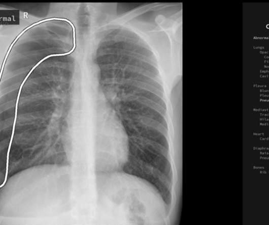
ChatGPT-4 outperformed human clinicians in determining pretest and post-test disease probability after a negative test result involving chest radiographs and mammograms, according to a research letter published December 11 in JAMA Network Open.
The rules also establish initial and continuing education requirements for limited-scope radiographers and radiologist assistants. We worked with the state to update the language that existed on equipment operation, education, clinical requirements, and credentialing," Aragon explained in the ARRT update. Read the full filing here.
Most Influential Radiology Researcher Erik Middlebrooks, MD, Mayo Clinic, Jacksonville, FL Erik Middlebrooks, MD. One of this year's nominations for Most Influential Radiology Researcher is Erik Middlebrooks, MD, of the Mayo Clinic in Jacksonville, FL.
Digital Radiography (DR) can help speed your imaging workflow, ease the workload on your staff, and reduce the dose to support a higher standard of care. Read on to learn four considerations when selecting solutions to make the transition from analog to digital radiography.
At RSNA 2023, look for AI-driven systems that radiographers can use to help make patient positioning faster and more precise, and bring consistency to the process, all of which help improve image quality and reduce the need for retakes. Even the most skilled radiographers can fail to get positioning just right.
Dynamic digital radiography (DDR) has shown for the first time that it can be used to automatically capture lung signal changes during forced breathing in patients with chronic obstructive pulmonary disease (COPD), according to a recent study. and Japanese developers wrote. and Japanese developers wrote. L and FEV1 of 0.68
In an open forum, Yi Xiang Tay, of Singapore University Hospital's radiography and diagnostic imaging department, shared his team's research. In addition, clinical decision support systems were the most evaluated mode of intervention, either integrated or standalone. For more coverage from ECR 2024, please visit our RADCast.
Initial results from currently ongoing clinical studies suggest that dark-field chest x-ray can be useful for detecting and quantification of pulmonary emphysema , and for assessing COVID-19 pneumonia , the authors wrote. The article was published March 7 in IEEE Transactions on Medical Imaging.
. | W3-SSMK08-4 | Room E450A A deep learning-based framework for automated screening of osteoporosis on lumbar spine plain radiographs shows potential as another way to opportunistically make use of imaging studies performed for other indications, according to this presentation.
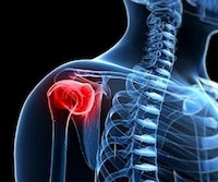
An AI algorithm may make dynamic digital radiography (DDR) more efficient by automatically measuring kinematics involved in certain shoulder injuries, according to a presentation delivered November 29 at RSNA. These images are then processed to visualize joints in motion.
So we were thinking and asking ourselves, ‘can nonradiologists benefit from AI and chest radiography analysis in this emergency unit set.’ ” Per year, LMU receives between 5,000 and 6,000 orders for chest radiographs for primary diagnosis from the emergency unit alone.
Researchers at the Icahn School of Medicine at Mount Sinai ("Icahn Mount Sinai") used Dynamic Digital Radiography (DDR) data, an X-ray imaging technology developed by Konica Minolta, to create their AI-powered technique that analyzes lung function. Chest radiography is typically acquired in the evaluation of pulmonary disorders.
Assessment of Claimant, Clinical, and Financial Characteristics of Teleradiology Medical Malpractice Cases. Commercially Available Chest Radiograph AI Tools for Detecting Airspace Disease, Pneumothorax, and Pleural Effusion. Generative Artificial Intelligence for Chest Radiograph Interpretation in the Emergency Department.
The technology figured prominently in five Minnies categories, including Hottest Clinical Procedure. Pickhardt believes using additional clinical data on CT imaging could save healthcare costs, especially when AI tools are used in parallel, suggesting it is a cost-effective or even cost-saving strategy for personalized or precision medicine.
Shamie Kumar describes how AI fits into a radiology clinical workflow and her perspective on how a clinicalradiographer could use this to learn from and enhance their skills. If the AI findings are seen in PACS, how many radiographers actually log into PACS after taking a scan or X-ray? Can Radiographers Up-Skill?
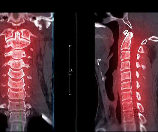
A highly accurate prediction rule for cervical spine injuries in children reduces the use of CT scans by over 50% without missing clinically significant injuries or increasing the use of unnecessary X-rays.
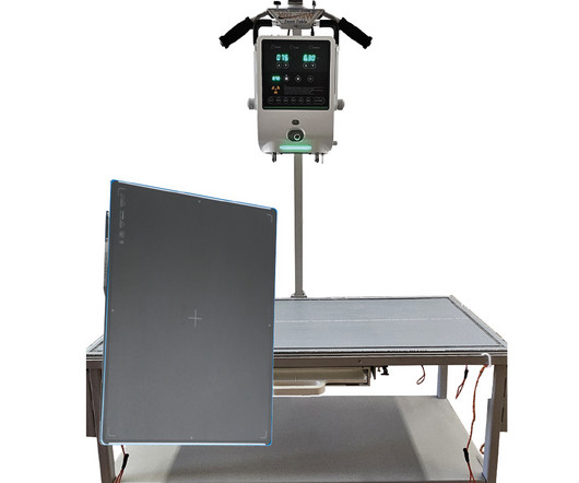
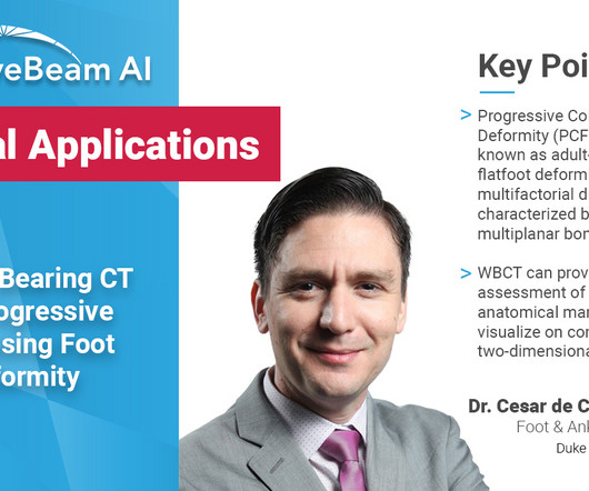
The following panelists of experts share their insights and experiences in the use of WBCT in clinical practice as well as the future direction of the modality. Whenever bilateral standing radiographs would have been needed, a WBCT was performed instead. How Frequently Do You Use It In Practice?
launch of its latest addition to its advanced digital radiography suites, the D-EVO Suite OTCx. The overhead tube crane system is designed to deliver consistent, efficient and comfortable patient exams, enhance technologist’s workflow, deliver clinical confidence, and help to keep imaging departments running smoothly.
Kim Mason Kim Mason, an Audit and Research Radiographer for Mid Yorkshire Teaching Hospitals Trust, talks about their role as well as the value of radiographer engagement in research activities and how to get involved. Hi, I’m Kim and I am an alternative-styled, funky-haired, septum-pierced, disabled Audit and Research Radiographer.
meets this need by developing clinical-grade AI models designed to improve capacity. Studies indicate that Harrison's AI for chest radiography can aid in the early detection of lung cancer, showing that over 32% of lung cancer cases could have been diagnosed soonerby an average of 16 months2. Co-Founder and Chief Executive Officer.
Konica Minolta Healthcare Americas and AI software developer Gleamer have entered into a strategic partnership in which Gleamer’s Bone View software will be added to Konica Minolta’s digital radiography (DR) product portfolio. The vendor says that this U.S.
Dynamic chest radiography (DCR) shows potential as a tool to investigate lung health in people with cystic fibrosis (CF), according to research published February 13 in Clinical Radiology. and colleagues. Yet FEV1 may not always reflect the severity of the airway obstruction, they noted.
Key Points: Currently plain radiographs are the standard method in diagnosing syndesmotic ankle injuries even though the distal tibiofibular joint cannot be assessed due to superposition of the osseous structures in the foot. Researchers used data from 76 NWBCT ankles (retroactive study) and 86 WBCT ankles.
Reading Time: 9 minutes read Carestream solutions help improve patient safety and clinical outcomes. A fundamental goal of radiographers is to complete an imaging exam that provides sufficient information for an accurate clinical diagnosis–and at the lowest possible dose.
Building on its century-long foundation of imaging research science, the company will showcase its diagnostic imaging solutions that help improve clinical outcomes, enhance the imaging experience for users and patients, and strengthen the financial position of healthcare facilities. “The
X-ray systems are a fundamental part of this, but the choice between a mobile X-ray system and a stationary one can greatly impact the workflow and efficiency of your clinic. Flexibility and Mobility Unlike stationary systems, mobile X-ray units can be transported to different locations within your clinic or even off-site for field work.
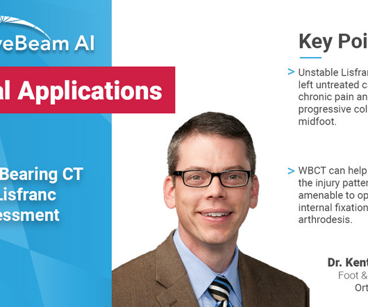
Diagnosis The diagnosis of Lisfranc injuries may be challenging on plain radiographs alone. Radiographs were indeterminate. MedShape and Curvebeam announce initiation of joint prospective clinical study. Identify subtle Lisfranc injuries by effectively differentiating between stable and unstable Lisfranc injuries 2. 2020.109419.
In the third blog of her series on AI and the radiographer, Shamie Kumar explores the impact on the radiographer when AI is integrated within an imaging modality. The question to explore in this blog is when AI is integrated within an imaging modality itself and how that may impact a radiographer.
Chest radiography is the most common medical imaging tool used in routine clinical practices to identify different disease findings. Augmento X-Ray is designed to significantly reduce radiologist workload and improve the quality of chest X-ray reporting. billion annual X-rays performed, 1.5
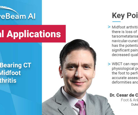
Diagnosis An accurate diagnosis and grading of the severity of osteoarthritic joints in the midfoot have been shown to be clinically relevant in treating the pathology early in its course and avoiding late-stage invasive procedures such as arthrodesis 3. Images demonstrated severe degeneration of the midfoot joints. Eur J Radiol.
The new imaging systems that will be on display include three new digital radiography (DR) suites, two new fluoroscopy systems, a 0.4T The Essentia SA is an ultra-compact straight arm system, designed for a wide range of standing, sitting and recumbent radiographic exams. MRI system and a 128- slice computed tomography (CT) system. “At
But how will AI in the workplace affect the radiographer and how does it differ from the red dot system radiographers are so familiar with? The Red Dot System Often one of the first courses a newly qualified radiographer attends is the red dot course. What does AI do that a radiographer doesn’t already?
With that said, it would be easy to theorize that more than anything else, that cost is the most crucial factor for clinics, hospitals, and imaging centers that are seeking X-Ray equipment solutions. But is that really the case? Who is Making the Purchases?
Key Points: Weight bearing CT (WBCT) can detect signs of osteoarthritis (OA), such as osteophytes, subchondral cysts, and joint space narrowing better than radiographs. The post WBCT and its Evolving Role in OA Research and Clinical Practice appeared first on CurveBeam AI. In addition, JSW does not decrease uniformly across the joint.
Project philosophical doughnut was an editorial project to tidy up, standardize and, most importantly, give clinical indications and purpose to our CT protocol articles (hence the wacky name Andrew chose and stuck with).
Carestream is here to help you navigate the turmoil, delivering solutions that help you create the best possible imaging experience for your staff and patients; improve clinical outcomes; and strengthen your financial health. Our DRX-LC Detector helps radiographers capture long-length images more effectively.
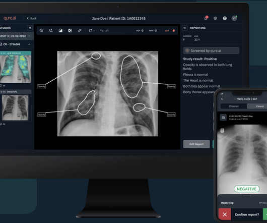
Qure’s chest X-ray based qXR-LN uses artificial intelligence to identify and localize lung nodules, marking another significant milestone for the organization, strengthening its standing as a pioneer in the realm of AI-powered advancements for plain film radiography and medical imaging. Monday, January 8, 2024 - 14:00
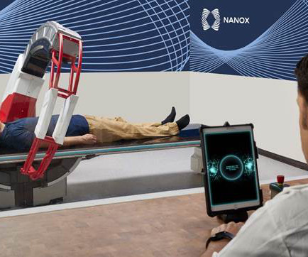
Nanox.ARC is a stationary X-ray system intended to produce tomographic images of the human musculoskeletal system adjunctive to conventional radiography on adult patients. Following this clearance, Nanox will continue to work with the FDA to pursue additional regulatory clearances and intends to expand clinical indications.
Reading Time: 9 minutes read Mid-cycle refresh can increase clinical, operational, and security benefits. Does it support new medical imaging software that can help improve clinical outcomes? Physicians have better diagnostic confidence when they can view radiographs in the manner most suitable to their preferences.
A weight bearing CT scan can: Provide an assessment of important anatomical markers of pronounced hindfoot deformity and peritalar subluxation (PTS), difficult to visualize on conventional two-dimensional radiographs 1. Allow for accurate evaluation of subtalar joint subluxation as well as sinus tarsi and subfibular impingement 2 .
We organize all of the trending information in your field so you don't have to. Join 5,000 users and stay up to date on the latest articles your peers are reading.
You know about us, now we want to get to know you!
Let's personalize your content
Let's get even more personalized
We recognize your account from another site in our network, please click 'Send Email' below to continue with verifying your account and setting a password.


Let's personalize your content