This site uses cookies to improve your experience. To help us insure we adhere to various privacy regulations, please select your country/region of residence. If you do not select a country, we will assume you are from the United States. Select your Cookie Settings or view our Privacy Policy and Terms of Use.
Cookie Settings
Cookies and similar technologies are used on this website for proper function of the website, for tracking performance analytics and for marketing purposes. We and some of our third-party providers may use cookie data for various purposes. Please review the cookie settings below and choose your preference.
Used for the proper function of the website
Used for monitoring website traffic and interactions
Cookie Settings
Cookies and similar technologies are used on this website for proper function of the website, for tracking performance analytics and for marketing purposes. We and some of our third-party providers may use cookie data for various purposes. Please review the cookie settings below and choose your preference.
Strictly Necessary: Used for the proper function of the website
Performance/Analytics: Used for monitoring website traffic and interactions
Philips and Vanderbilt assessed 13 diagnostic imaging devices including MR , CT , ultrasound and X-ray , which account for an estimated 12,000 patient scans per month, and found that they emit the CO₂ equivalent of 1,000 gas cars driven for one year.
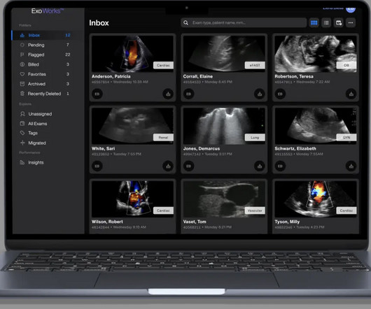
Exo Works Connect streamlines ultrasound exam documentation, enables real-time collaboration and feedback, simplifies quality assurance and tracks user proficiency—all accessible from a smartphone, tablet or computer. Exo Works Connect accommodates users of all levels and the solution can be onboarded quickly and efficiently.”
PACS – PictureArchiving and CommunicationSystem; a system involved in acquiring the medical images, transmission, viewing, storage, and retrieval of same images. The fundamental parts of PACS are – imaging acquisition, display workstations, archive servers. All this is possible because of PACS.
This blog post explores how the seamless incorporation of teleradiology into healthcare systems leads to improved diagnostics, streamlined workflows, and ultimately, enhanced patient care. Explore how systems are designed to handle data from modalities such as X-ray, CT, MRI, and ultrasound, providing a comprehensive diagnostic overview.
Teleradiology operates by leveraging digital communication technologies to transmit medical images, such as X-rays, MRIs, CT scans, and ultrasounds, from one location to another for interpretation and diagnosis. Remote Interpretation: Radiologists, often located at a different site or working remotely, receive these images.
Spotlight key technological breakthroughs, including computed tomography (CT), magnetic resonance imaging (MRI), and ultrasound, revolutionizing the diagnostic panorama.
CPACS stands for Cardiovascular PictureArchive and CommunicationSystem. It is a medical imaging system used to store, manage, and share cardiology images. Cardiologists use CPACS systems to view, diagnose, and treat heart conditions. How Does CPACS Work?
In the field of cardiology, medical imaging systems play an integral role in providing effective care. As technology advances, these systems continue to evolve to meet the changing needs of healthcare organizations. CPACS and CVIS, both healthcare information systems, are used to store, manage, archive, and communicate medical images.
The theoretical basis for ultrasound physics has been around since 1794, but it wasn’t until 1942, when Dr Karl Theodore Dussik in Austria transmitted an ultrasound beam through a human skull to view the brain, that ultrasound was first used in medicine. (13) This was a defining publication in the field of medical ultrasound. (14)
One such technology is the cloud-based picturearchiving and communicationsystems or PACS. This system offers unmatched efficiency., This includes tools like MR, CT and ultrasound, positron emission tomography. Large radiology companies even have malpractice insurance to save you from costly legal actions.
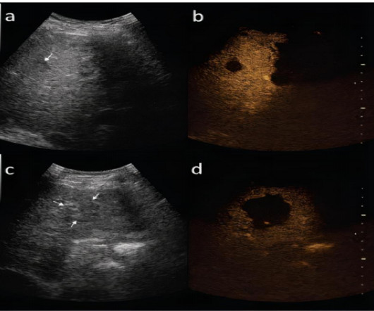
DIAGNOSIS OF HEPATOCELLULAR CARCINOMA IMAGING MODALITIES ULTRASOUND: Small focal HCC appears hypoechoic compared with normal liver. All image data were transmitted directly to our picturearchiving and communicationsystem. Tumour thrombus may be visible.(8) Tumour thrombus may be visible.(8)
We organize all of the trending information in your field so you don't have to. Join 5,000 users and stay up to date on the latest articles your peers are reading.
You know about us, now we want to get to know you!
Let's personalize your content
Let's get even more personalized
We recognize your account from another site in our network, please click 'Send Email' below to continue with verifying your account and setting a password.


Let's personalize your content