This site uses cookies to improve your experience. To help us insure we adhere to various privacy regulations, please select your country/region of residence. If you do not select a country, we will assume you are from the United States. Select your Cookie Settings or view our Privacy Policy and Terms of Use.
Cookie Settings
Cookies and similar technologies are used on this website for proper function of the website, for tracking performance analytics and for marketing purposes. We and some of our third-party providers may use cookie data for various purposes. Please review the cookie settings below and choose your preference.
Used for the proper function of the website
Used for monitoring website traffic and interactions
Cookie Settings
Cookies and similar technologies are used on this website for proper function of the website, for tracking performance analytics and for marketing purposes. We and some of our third-party providers may use cookie data for various purposes. Please review the cookie settings below and choose your preference.
Strictly Necessary: Used for the proper function of the website
Performance/Analytics: Used for monitoring website traffic and interactions
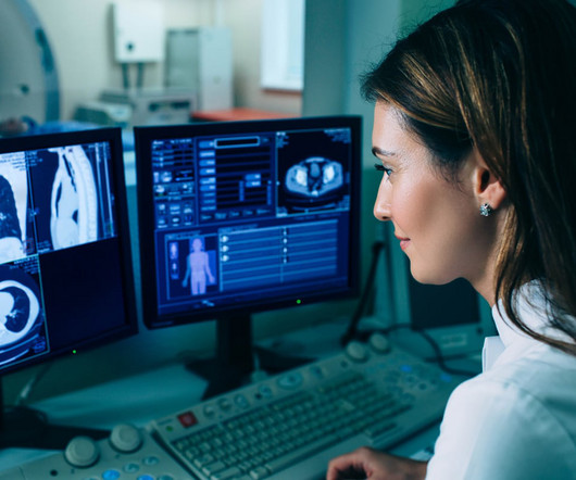
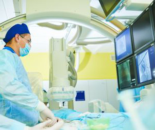
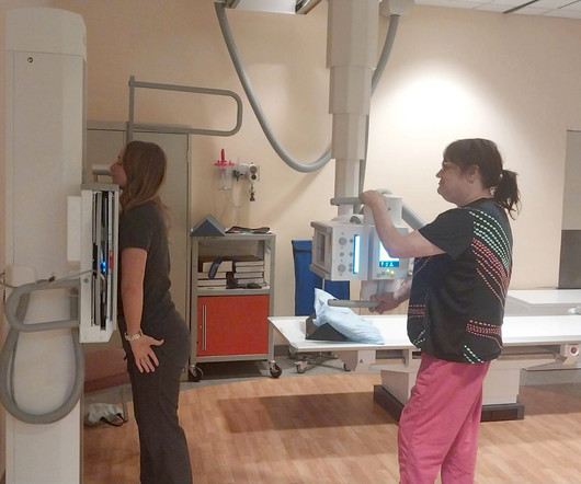
If you’re looking for diagnostic imaging services in the El Paso region, contact us online or call (915) 225-2480 to schedule an appointment today. MagneticResonanceImaging (MRI) This type of diagnostic imaging, commonly known as MRI , uses powerful magnetic fields and radio waves to create detailed images of the internal body.
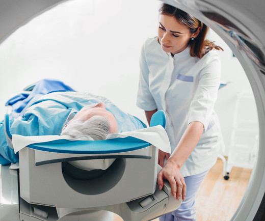
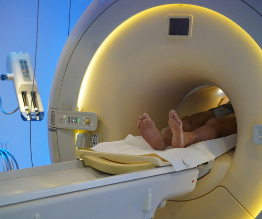
Computedtomography (CT scans) plays a critical role in diagnosing and monitoring a wide range of medical conditions. At Professional Radiology , we use state-of-the-art medical machinery to successfully provide patients in El Paso with accurate imaging services.
Computedtomography (CT scans) plays a critical role in diagnosing and monitoring a wide range of medical conditions. At Professional Radiology , we use state-of-the-art medical machinery to successfully provide patients in El Paso with accurate imaging services.
The Importance of Early Detection Through Imaging Early detection of cancer through imaging allows for interventions at stages when treatment is most effective. Teleradiology: Bridging the Gap in Underserved Areas Teleradiology plays a pivotal role in expediting cancer diagnoses, particularly in rural or underserved regions.
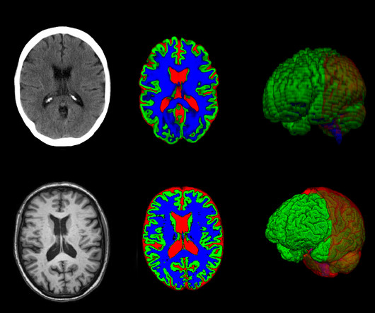
milla1cf Fri, 10/20/2023 - 18:29 October 20, 2023 — In certain cases, a new method can provide as much information from brain images taken with computedtomography (CT) as images captured with magneticresonanceimaging (MRI).
milla1cf Thu, 01/04/2024 - 10:47 January 4, 2024 — Diagnosing cancer today involves using chemical “contrast agents” to improve the accuracy of medical imaging processes such as X-rays as well as computedtomography (CT) and magneticresonanceimaging (MRI) scans.
Diagnostic imaging is an important tool used every day in healthcare to assist doctors in making the most informed decisions for their patients. Over the years, advancements in diagnostic imaging have greatly increased patients’ overall care, quality of life, and outcome when diagnosed with certain conditions.
Key Points: Cone Beam CT (CBCT) is superior in assessing bony structures compared to magneticresonanceimaging (MRI) In this study, there was a 40% rate of discrepancy when grading knee subchondral insufficiency fractures on CBCT vs. MRI, with MRI frequently underestimating damage of the subchondral bone plate while overestimating lesion size.
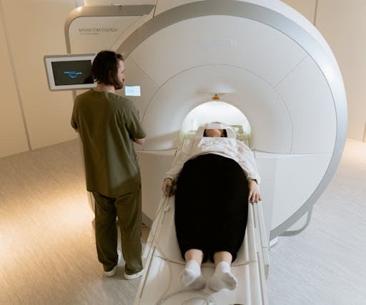
MagneticResonanceImaging (MRI) MRI stands for MagneticResonanceImaging. Instead, they employ a powerful magnetic field along with radio waves to produce detailed images of organs and tissues. Joint and Muscle Disorders: It is excellent for imaging knee and shoulder injuries.
As seen with other disease patterns, many operators subsequently obtain cross-sectional or dynamic imaging for further evaluation. Computedtomography (CT) angiography, which has a 100% sensitivity for diagnosing this entity, is commonly used (1).
The approval expands upon Bayer's focus on breast imaging, with a portfolio that also includes Gadavist (gadobutrol) injection, a gadolinium-based contrast agent approved for use with MRI ( MagneticResonanceImaging ) to assess the presence and extent of malignant breast disease in adult patients. for use in CEM.
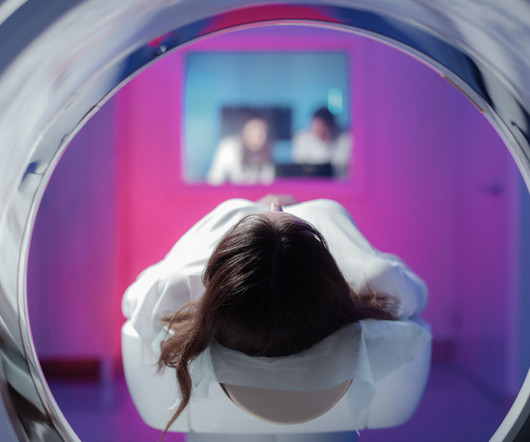
Magneticresonanceimaging (MRI) is used to help diagnose and treat various medical conditions. At Intermountain Medical Imaging , we rely on a variety of MRI options that offer a wider opening, helping us deliver high-quality care to clients and medical providers alike. MRIs rely on large magnets and radio waves.
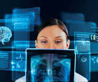
Medical imaging is crucial in diagnosing and treating various medical conditions. These technologies have transformed the medical field from X-rays to MagneticResonanceImaging (MRI) and ComputedTomography ( CT ) scans.
Subspecialized Expertise Consider the wide-ranging scope of your medical imaging needs. At various times, you may need to rely on an expert to interpret images produced by X-ray, magneticresonanceimaging (MRI), computedtomography (CT), positron emission tomography/computedtomography (PET/CT), or another modality.
Magneticresonanceimaging (MRI) is an essential tool that healthcare providers use to diagnose and treat various conditions. It provides highly detailed images of what is going on inside the body and how multiple systems work with each other.
Kidney cancer can be a life-threatening condition if it is not diagnosed and treated early. Kidney cancer is often diagnosed using imaging tests that show visualizations of the kidneys as well as any abnormal growths of cancer cells within them. This is delivered intravenously.
In 2011, a large study examined the use of x-rays and other radiation imaging on children—they estimated that the average child would get more than seven radiation scans by the age of 18. No doubt, then, that the role of a pediatric radiologist is important in accurately diagnosing and treating diseases and conditions in children.
Beyond Conventional Radiography: Advanced Imaging Modalities: Digital technology expanded the scope of X-ray imaging beyond traditional radiography. Computedtomography (CT) and magneticresonanceimaging (MRI) emerged as advanced modalities, providing detailed 3D images of the body and opening new possibilities for diagnosis.
Only 21% of extremely disadvantaged zip codes had access to computedtomography (CT) facilities as compared to 32% of extremely advantaged zip codes. As for magneticresonanceimaging (MRI), just 19% of extremely disadvantaged zip codes had access as compared to 32% of extremely advantaged.
As radiology departments proliferated in hospitals, X-rays became indispensable for diagnosing a wide range of conditions. Traditional film-based X-rays gave way to digital radiography (DR) and computed radiography (CR). Advanced Imaging Modalities: X-ray technology has expanded beyond conventional radiography.
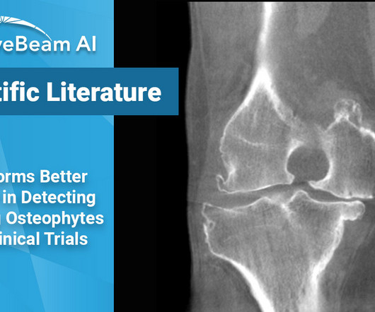
Key Points: While magneticresonanceimaging (MRI) adequately detects the size and presence of osteophytes (OPs) in the medial tibio-femoral compartment, it underestimates OPs in all other knee compartments of osteoarthritic patients. Clinical trials will often use MRI to assess and grade OPs in arthritic knees.
Medical imaging is used to find, diagnose, monitor, and even treat different medical conditions or injury. Each piece of technology encompassed in medical imaging focuses on a different area or system of the body. Medical imaging, as a practice, has been around since the last 19th century.
When it comes to accurate diagnoses and effective patient care, getting a second opinion on imaging results can make all the difference. Types of Imaging Studies That Often Need a Second Opinion Not all imaging studies are straightforward, and some require deeper insight to reach a definitive diagnosis.
of all diagnosed fractures. With over 20% of Lisfranc fractures missed upon presentation, it is important to diagnose these injuries promptly, as lack of diagnosis could lead to future foot deformities, midfoot arthritis, pain, chronic instability, and disability [1,5]. J Foot Ankle Res. 2023;16(1):9. Published 2023 Mar 1.
Doctors use imaging tests to see inside a patient’s body and diagnose their illnesses and injuries. There are many different types of imaging tests, each with their own method of generating a photo of the inside of the human body. A doctor will often use a diagnostic imaging test to check patients for signs of cancer.
It is a medical imaging system used to store, manage, and share cardiology images. Cardiologists use CPACS systems to view, diagnose, and treat heart conditions. Like PACS , CPACS systems typically use DICOM (Digital Imaging and Communications in Medicine) standards to store and transmit images.
This standard has revolutionized the radiology industry, encompassing many imaging modalities such as X-rays, computedtomography (CT), magneticresonanceimaging (MRI), ultrasound, nuclear medicine, PET scans, etc. They can enhance image quality, generate additional data, take measurements, and more.
A CVIS (or CIS) is a specialized software platform designed to manage, store, and analyze clinical information related to diagnosing, treating, and managing cardiovascular diseases. The system can consolidate the results of the current diagnostic tests, such as ECG, lab tests, and imaging studies, and display them in an organized manner.
Computer aided diagnostic systems are already in use throughout radiology and can accurately diagnose breast cancer with a higher degree of accuracy than their human counterparts (2) and this use will only increase. New nanotechnologies may soon allow targeted and molecular imaging of tumours and other conditions.
Tomography originates from the Greek words ‘tomos’, meaning ‘slice’ or ‘section’, and ‘graphia’, meaning ‘description of’. In the 1960s computers were increasingly available and more powerful and in 1971, the first computedtomography (CT) scan was performed on a patient.
Key Points: Imaging modalities such as plain radiographs (X-Ray), computedtomography (CT), and magneticresonanceimaging (MRI), dont have the diagnostic accuracy needed to detect syndesmotic widening or subtle instability. Specimens were mounted in a frame that allowed simulated axial weight bearing.
Magneticresonanceimaging and computedtomography in emergency assessment of patients with suspected acute stroke: a prospective comparison. Contrast enhancement and contrast extravasation on computedtomography after intra-arterial thrombolysis in patients with acute ischemic stroke. Mancall EL.
We organize all of the trending information in your field so you don't have to. Join 5,000 users and stay up to date on the latest articles your peers are reading.
You know about us, now we want to get to know you!
Let's personalize your content
Let's get even more personalized
We recognize your account from another site in our network, please click 'Send Email' below to continue with verifying your account and setting a password.


Let's personalize your content