This site uses cookies to improve your experience. To help us insure we adhere to various privacy regulations, please select your country/region of residence. If you do not select a country, we will assume you are from the United States. Select your Cookie Settings or view our Privacy Policy and Terms of Use.
Cookie Settings
Cookies and similar technologies are used on this website for proper function of the website, for tracking performance analytics and for marketing purposes. We and some of our third-party providers may use cookie data for various purposes. Please review the cookie settings below and choose your preference.
Used for the proper function of the website
Used for monitoring website traffic and interactions
Cookie Settings
Cookies and similar technologies are used on this website for proper function of the website, for tracking performance analytics and for marketing purposes. We and some of our third-party providers may use cookie data for various purposes. Please review the cookie settings below and choose your preference.
Strictly Necessary: Used for the proper function of the website
Performance/Analytics: Used for monitoring website traffic and interactions
DiagnosticImaging is a great tool for your medical professional to use to detect issues sooner rather than later. There are several types of diagnosticimaging available today; each one used to visualize the internal structures of the body to assist doctors in diagnosis and treating various diseases and medical conditions.
When reviewing radiographs, computedtomography (CT) scans or magneticresonanceimaging (MRI) scans, do you still turn to mnemonics every now and then to jog your short-term memory?
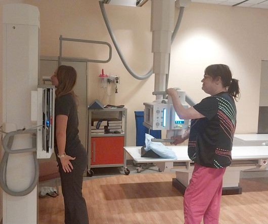
Computedtomography (CT scans) plays a critical role in diagnosing and monitoring a wide range of medical conditions. At Professional Radiology , we use state-of-the-art medical machinery to successfully provide patients in El Paso with accurate imaging services.
Diagnosticimaging is an important tool used every day in healthcare to assist doctors in making the most informed decisions for their patients. Over the years, advancements in diagnosticimaging have greatly increased patients’ overall care, quality of life, and outcome when diagnosed with certain conditions.
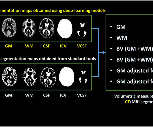
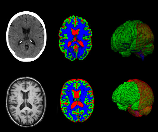
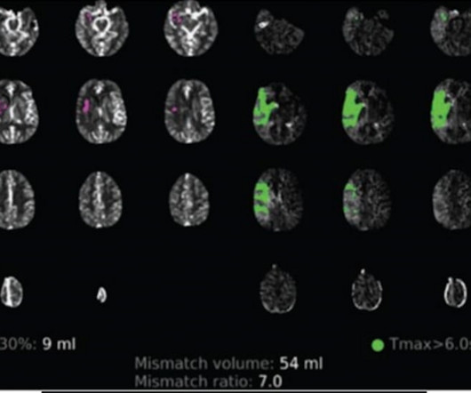
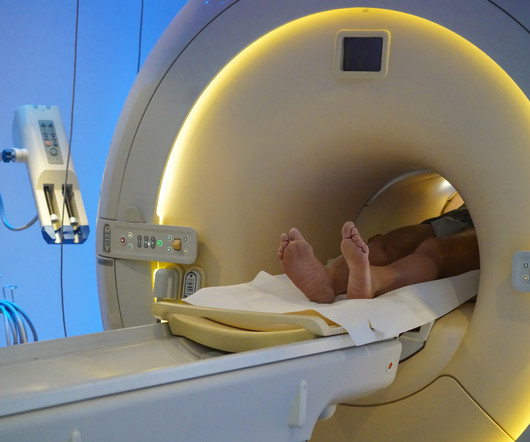
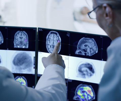
In certain cases, a new method can provide as much information from brain images taken with computedtomography as images captured with magneticresonanceimaging.
In certain cases, a new method can provide as much information from brain images taken with computedtomography (CT) as images captured with magneticresonanceimaging (MRI).
Campione also joins the Blue Earth Diagnostics Ltd. Board of Directors and will serve as Vice Chair of the Blue Earth Diagnostics Inc. Board of Directors and will serve as Vice Chair of the Blue Earth Diagnostics Inc. Blue Earth Diagnostics, an indirect subsidiary of Bracco Imaging S.p.A., Bracco Imaging S.p.A.,
The Radiological Society of North America’s (RSNA) 2024 annual meeting showcased significant advancements in medical imaging, including artificial intelligence (AI), innovations in computedtomography (CT) and magneticresonanceimaging (MRI), and strategies to address the ongoing radiology staffing shortage.
milla1cf Fri, 10/20/2023 - 18:29 October 20, 2023 — In certain cases, a new method can provide as much information from brain images taken with computedtomography (CT) as images captured with magneticresonanceimaging (MRI).
Computedtomography (CT scans) plays a critical role in diagnosing and monitoring a wide range of medical conditions. At Professional Radiology , we use state-of-the-art medical machinery to successfully provide patients in El Paso with accurate imaging services.
Key Points: Cone Beam CT (CBCT) is superior in assessing bony structures compared to magneticresonanceimaging (MRI) In this study, there was a 40% rate of discrepancy when grading knee subchondral insufficiency fractures on CBCT vs. MRI, with MRI frequently underestimating damage of the subchondral bone plate while overestimating lesion size.
milla1cf Thu, 01/04/2024 - 10:47 January 4, 2024 — Diagnosing cancer today involves using chemical “contrast agents” to improve the accuracy of medical imaging processes such as X-rays as well as computedtomography (CT) and magneticresonanceimaging (MRI) scans.
The Importance of Early Detection Through Imaging Early detection of cancer through imaging allows for interventions at stages when treatment is most effective. These benefits collectively enhance the overall performance of healthcare facilities by streamlining operations and ensuring high-quality diagnostic services.
Unvaccinated people with COVID-19 who undergo angiographic reperfusion after acute ischemic stroke may have a greater than fivefold risk of continued infarct growth in comparison to unvaccinated people without COVID-19, according to computedtomography perfusion (CTP) and magneticresonanceimaging (MRI) findings from a recently published study.
MagneticResonanceImaging (MRI) MRI stands for MagneticResonanceImaging. Instead, they employ a powerful magnetic field along with radio waves to produce detailed images of organs and tissues. Joint and Muscle Disorders: It is excellent for imaging knee and shoulder injuries.
For the third year in a row, a Capitol Imaging Services affiliate has been designate as the best of the best! Our Southeast Louisiana network member, DiagnosticImaging Services (DIS), was chosen by the readers of EDGE Magazine published in St. Tammany Parish.
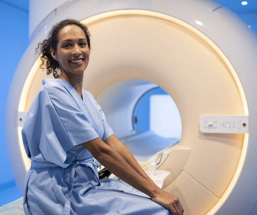
Magneticresonanceimaging (MRI) is used to help diagnose and treat various medical conditions. At Intermountain Medical Imaging , we rely on a variety of MRI options that offer a wider opening, helping us deliver high-quality care to clients and medical providers alike. MRIs rely on large magnets and radio waves.
Essential in a wide variety of health care settings, medical imaging is a key diagnostic tool for many conditions and integral for both monitoring treatment and predicting outcomes. Subspecialized Expertise Consider the wide-ranging scope of your medical imaging needs.
Discuss the initial applications of radiography and its profound impact on the landscape of medical diagnostics. Technological Metamorphosis: From Analog to Digital Prowess Chart the transition from analog radiology to the epoch of digital imaging technologies.
ROCC connects imaging experts at a Command Center with technologists at scanning locations across an organization. According to Gupta, Philips Image Orchestrator has been well received by diagnosticimaging centers. We are also seeing our customers [using ROCC] for virtually assisted scanning as well,” Phanse said.
Magneticresonanceimaging (MRI) is an essential tool that healthcare providers use to diagnose and treat various conditions. It provides highly detailed images of what is going on inside the body and how multiple systems work with each other.
Section 2: Extraneous Imaging Defined: Define extraneous imaging in the context of trauma radiology, exploring the various imaging modalities beyond conventional radiography and computedtomography (CT) scans.
Medical imaging is crucial in diagnosing and treating various medical conditions. These technologies have transformed the medical field from X-rays to MagneticResonanceImaging (MRI) and ComputedTomography ( CT ) scans.
The Digital Advantage: Image Clarity and Efficiency: Digital radiography brought newfound clarity to X-ray images, offering healthcare professionals the ability to zoom in, adjust contrast, and enhance details. The digital advantage also accelerated the diagnostic process, leading to faster, more efficient patient care.
The Birth of X-ray Technology: At the end of the 19th century, Wilhelm Conrad Roentgen’s discovery of X-rays opened up new possibilities in medical diagnostics. Advanced Imaging Modalities: X-ray technology has expanded beyond conventional radiography.
Types of Imaging Services that Detect Kidney Cancer Computedtomography , commonly referred to as a CT scan, creates images of the body’s internal structures and can provide detailed information regarding the size and shape of a tumor in the kidney. This is delivered intravenously.
The role of a pediatric radiologist involves the following: Interpreting Imaging Studies: Pediatric radiologists review and interpret various imaging studies such as X-rays, ultrasounds, computedtomography (CT) scans, magneticresonanceimaging (MRI), and nuclear medicine scans.
To meet that goal, we had to get the best possible equipment and that’s why we turned to Fujifilm Healthcare Americas Corporation for the very latest in diagnosticimaging systems.” Only 21% of extremely disadvantaged zip codes had access to computedtomography (CT) facilities as compared to 32% of extremely advantaged zip codes.
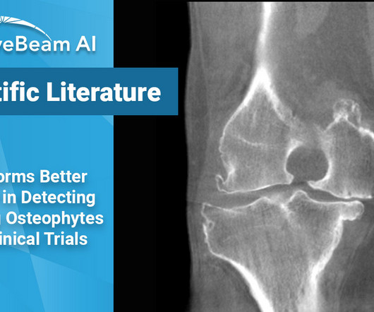
Key Points: While magneticresonanceimaging (MRI) adequately detects the size and presence of osteophytes (OPs) in the medial tibio-femoral compartment, it underestimates OPs in all other knee compartments of osteoarthritic patients. Dr. Roemer et al. When it came to OP size, the PFJ MRI underestimated the size 21% of the time.
Whether you’re looking for a second set of eyes for complex cases or want to ensure the highest level of diagnostic accuracy, our team of board-certified radiologists—with subspecialties in areas such as neuroradiology, musculoskeletal imaging, and oncology—are ready to assist. Why Choose a Teleradiology Partner for Second Opinions?
Doctors use imaging tests to see inside a patient’s body and diagnose their illnesses and injuries. There are many different types of imaging tests, each with their own method of generating a photo of the inside of the human body. A doctor will often use a diagnosticimaging test to check patients for signs of cancer.
christine.book Thu, 05/02/2024 - 12:01 May 2, 2024 — GE HealthCare has announced a new radiation therapy computedtomography (CT) solution with innovative hardware and software solutions to help increase imaging accuracy.
A computedtomography scan will better assist with diagnosis and help with planning if surgery is necessary [3]. Magneticresonanceimaging will help to evaluate ligamentous injury and provides a 94% predictive value for diagnosing Lisfranc injury [5]. Chen C, Jiang J, Wang C, Zou J, Shi Z, Yang Y. 2023;16(1):9.
I believe that the first main difference seen in the radiologists role in 2040 will be the change that artificial intelligence (AI) will have made on the diagnostic role that radiologists currently perform. Nanotechnology Development and Utilization: A Primer for Diagnostic and Interventional Radiologists. Hough, Andrew.
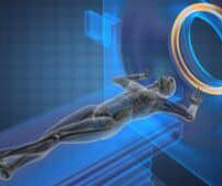
MRI , or MagneticResonanceImaging, is diagnostic technology that uses a powerful magnetic field and computer-generated radio waves to create detailed, 3D images of the organs and tissues of the body that can be viewed from many different angles.
3D medical imaging transforms diagnostics and treatment, enhancing precision, patient education, and enabling AI-driven analysis and immersive experiences. The post A Dive into the World of 3D Medical Imaging appeared first on Open Medscience.
In the ever-evolving landscape of modern medicine, technology plays a crucial role in transforming patient care, enhancing diagnostic accuracy, and streamlining workflows. It is a medical imaging system used to store, manage, and share cardiology images. Cardiac CT is a type of computedtomography used to image the heart.
Cardiovascular Information Systems (CVIS) streamline clinical data management, diagnostics, and treatment for heart-related conditions, enhancing patient care. Before surgery, patients undergo various diagnosticimaging tests, such as echocardiograms, CT scans, and MRIs. Consider the case of cardiac surgery.
Tomography originates from the Greek words ‘tomos’, meaning ‘slice’ or ‘section’, and ‘graphia’, meaning ‘description of’. In the 1960s computers were increasingly available and more powerful and in 1971, the first computedtomography (CT) scan was performed on a patient. Computer diagnosis of primary bone tumor.
This standard has revolutionized the radiology industry, encompassing many imaging modalities such as X-rays, computedtomography (CT), magneticresonanceimaging (MRI), ultrasound, nuclear medicine, PET scans, etc.
Can AI and machine learning–driven care meet these standards — ones that we demand from a novel therapeutic intervention or laboratory-based diagnostic test — or do we need to have a unique set of standards for this type of intervention? 3 tables, 1 figure, no images 6. 3 figures 4. Lee BC, Tsai HH, Liu CJ, et al. 2023;54(4):1046-1055.
Key Points: Imaging modalities such as plain radiographs (X-Ray), computedtomography (CT), and magneticresonanceimaging (MRI), dont have the diagnostic accuracy needed to detect syndesmotic widening or subtle instability. To read the full study click here.
Magneticresonanceimaging and computedtomography in emergency assessment of patients with suspected acute stroke: a prospective comparison. Contrast enhancement and contrast extravasation on computedtomography after intra-arterial thrombolysis in patients with acute ischemic stroke.
We organize all of the trending information in your field so you don't have to. Join 5,000 users and stay up to date on the latest articles your peers are reading.
You know about us, now we want to get to know you!
Let's personalize your content
Let's get even more personalized
We recognize your account from another site in our network, please click 'Send Email' below to continue with verifying your account and setting a password.


Let's personalize your content