Remove Computed Tomography Remove Global Radiology Remove Presentation
article thumbnail

Lens Subluxation

Global Radiology CME

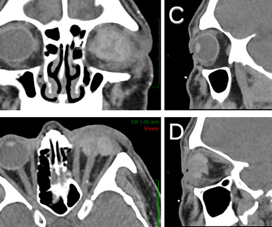
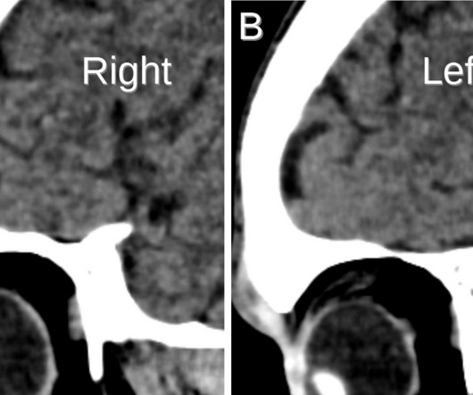
Patients with ectopia lentis typically present with visual acuity problems but can also present with eye pain if secondary to trauma [2]. Computed tomography is also an alternative method for lens subluxation which again can show deviation of the lens (Figs. Acta Med Port. 2020;33(10):692. doi: 10.20344/amp.12418

article thumbnail

Ruptured Globe

Global Radiology CME

1 Imaging: Computed tomography (CT) is the recommended imaging modality for evaluating orbital trauma. Thin-section axial CT scans followed by multiplanar reformations can visualize ruptured globe contour, which can present as a “mushroom” (Figs. 1-2) or “flat tire” shape. 7 Prognosis: Ruptured globes maintain a poor prognosis.

Insiders

Sign Up for our Newsletter

This site is protected by reCAPTCHA and the Google Privacy Policy and Terms of Service apply.

Trending Sources

article thumbnail

Lisfranc Fracture Dislocation

Global Radiology CME

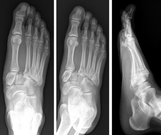
With over 20% of Lisfranc fractures missed upon presentation, it is important to diagnose these injuries promptly, as lack of diagnosis could lead to future foot deformities, midfoot arthritis, pain, chronic instability, and disability [1,5]. She is now pursuing a career in Diagnostic Radiology with interests in Breast imaging.

article thumbnail

Acute Infective Vasitis

Global Radiology CME

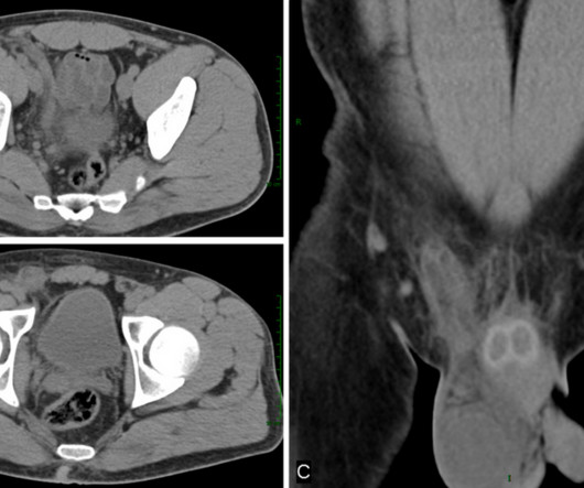
Vasitis often presents as painful swelling in the groin with a palpable mass in the scrotal region (3). It can also present with leukocytosis, fever, and right lower quadrant pain as seen in this patient (3). Vasitis: clinical and ultrasound confusion with inguinal hernia clarified by computed tomography. BMC Urology.

article thumbnail

Emphysematous Pyelonephritis

Global Radiology CME

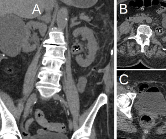
Like pyelonephritis, patients with EPN often present with fever, abdominal or flank pain, and costovertebral angle tenderness. KUB indicates kidneys, ureter, and bladder (plain abdominal radiograph); CT, computed tomography; and PCD, percutaneous catheter drainage. Pyelonephritis: Radiologic-Pathologic Review.