This site uses cookies to improve your experience. To help us insure we adhere to various privacy regulations, please select your country/region of residence. If you do not select a country, we will assume you are from the United States. Select your Cookie Settings or view our Privacy Policy and Terms of Use.
Cookie Settings
Cookies and similar technologies are used on this website for proper function of the website, for tracking performance analytics and for marketing purposes. We and some of our third-party providers may use cookie data for various purposes. Please review the cookie settings below and choose your preference.
Used for the proper function of the website
Used for monitoring website traffic and interactions
Cookie Settings
Cookies and similar technologies are used on this website for proper function of the website, for tracking performance analytics and for marketing purposes. We and some of our third-party providers may use cookie data for various purposes. Please review the cookie settings below and choose your preference.
Strictly Necessary: Used for the proper function of the website
Performance/Analytics: Used for monitoring website traffic and interactions
Computedtomography is also an alternative method for lens subluxation which again can show deviation of the lens (Figs. 2015.01.004 Sai Kilaru is a medical student at Central Michigan University College of Medicine and plans to pursue a residency in diagnostic radiology. J Emerg Med. 2015;48(6):e135-e137.
1 Imaging: Computedtomography (CT) is the recommended imaging modality for evaluating orbital trauma. Accuracy of ComputedTomography Imaging Criteria in the Diagnosis of Adult Open Globe Injuries by Neuroradiology and Ophthalmology. 1-2) or “flat tire” shape. Clin Ophthalmol. 2022;16:2545-2559. Acad Emerg Med.
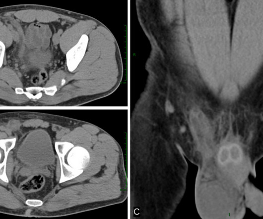
Vasitis: clinical and ultrasound confusion with inguinal hernia clarified by computedtomography. Clinical Radiology. Rice, MD is the president of GlobalRadiology CME and is a radiologist with Cape Radiology Group. Can Urol Assoc J. 2011;5(4):E74-E76. doi:10.5489/cuaj.10116 2011;66(5):475-477.
More sophisticated imaging, such as computedtomography or magnetic resonance imaging, should be obtained if plain film is unrevealing but there is high suspicion of fracture. He is vice-president of his school’s radiology interest group and a member of Rad Boot Camp. Diagram showing the types of fracture of the cuboid [1].
A computedtomography scan will better assist with diagnosis and help with planning if surgery is necessary [3]. Utility of weight-bearing radiographs compared to computedtomography scan for the diagnosis of subtle Lisfranc injuries in the emergency setting. J Foot Ankle Res. 2023;16(1):9. Published 2023 Mar 1.
KUB indicates kidneys, ureter, and bladder (plain abdominal radiograph); CT, computedtomography; and PCD, percutaneous catheter drainage. In severe cases or patients who do not respond to PCD, treatment with nephrectomy can lead to clinical and radiological improvement (Fig. doi: 10.1148/radiology.198.2.8596845
Computedtomography (CT) could be used to determine the capsule’s location if it is difficult to do so via x-ray. At UMKC, Eric has made significant contributions to the Radiology Interest Group, serving in various executive roles and currently as the interventional radiology chair.
We organize all of the trending information in your field so you don't have to. Join 5,000 users and stay up to date on the latest articles your peers are reading.
You know about us, now we want to get to know you!
Let's personalize your content
Let's get even more personalized
We recognize your account from another site in our network, please click 'Send Email' below to continue with verifying your account and setting a password.


Let's personalize your content