This site uses cookies to improve your experience. To help us insure we adhere to various privacy regulations, please select your country/region of residence. If you do not select a country, we will assume you are from the United States. Select your Cookie Settings or view our Privacy Policy and Terms of Use.
Cookie Settings
Cookies and similar technologies are used on this website for proper function of the website, for tracking performance analytics and for marketing purposes. We and some of our third-party providers may use cookie data for various purposes. Please review the cookie settings below and choose your preference.
Used for the proper function of the website
Used for monitoring website traffic and interactions
Cookie Settings
Cookies and similar technologies are used on this website for proper function of the website, for tracking performance analytics and for marketing purposes. We and some of our third-party providers may use cookie data for various purposes. Please review the cookie settings below and choose your preference.
Strictly Necessary: Used for the proper function of the website
Performance/Analytics: Used for monitoring website traffic and interactions
Imaging was so important [for cardiac indications], that I decided to become a radiologist," he said. Together with our radiographers, I learned to scan cardiac patients and learned special anatomy from pediatric cardiologists and pediatric cardiac surgeons." years of follow-up compared with CT imaging (2.1% But he persevered.
Mitigating radiology workforce strain requires revisioning the specialty and adding new roles to manage imaging demand, according to a presentation delivered September 12 at the International Society for ComputedTomography (ISCT) 2024 meeting in Boston.
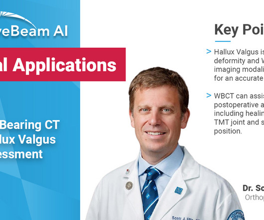
Diagnosis While radiographs are typically sufficient to make the diagnosis, WBCT scans may be useful to plan surgical treatment. Accurately assess sesamoid position as plain radiographs cannot determine whether the sesamoids have been reduced within their grooves 5. Accurately assess healing in the 1st TMT joint 4. 10.30795/jfootankle.2022.v16.1674.
1 Imaging: Computedtomography (CT) is the recommended imagingmodality for evaluating orbital trauma. Imaging of orbital trauma. Radiographics. Accuracy of ComputedTomographyImaging Criteria in the Diagnosis of Adult Open Globe Injuries by Neuroradiology and Ophthalmology.
after seeing the image. (2) Photoprint from radiograph by W.K. 3) In the early twentieth century, it was a common goal for investigators to try to find a way to separate the superimposed shadows that were recorded when a complex structure was shown on a radiograph. (3) 3) This is what is known as tomography.
Key Points: Imagingmodalities such as plain radiographs (X-Ray), computedtomography (CT), and magnetic resonance imaging (MRI), dont have the diagnostic accuracy needed to detect syndesmotic widening or subtle instability.
3] To identify a causative vascular lesion, which may or may not be amenable or contraindicatory to thrombolysis Non-Contrast Head CT NCCT is usually the first imagingmodality obtained in the acute evaluation for stroke. Radiographics. Perfusion computedtomography to assist decision making for stroke thrombolysis.
We organize all of the trending information in your field so you don't have to. Join 5,000 users and stay up to date on the latest articles your peers are reading.
You know about us, now we want to get to know you!
Let's personalize your content
Let's get even more personalized
We recognize your account from another site in our network, please click 'Send Email' below to continue with verifying your account and setting a password.


Let's personalize your content