This site uses cookies to improve your experience. To help us insure we adhere to various privacy regulations, please select your country/region of residence. If you do not select a country, we will assume you are from the United States. Select your Cookie Settings or view our Privacy Policy and Terms of Use.
Cookie Settings
Cookies and similar technologies are used on this website for proper function of the website, for tracking performance analytics and for marketing purposes. We and some of our third-party providers may use cookie data for various purposes. Please review the cookie settings below and choose your preference.
Used for the proper function of the website
Used for monitoring website traffic and interactions
Cookie Settings
Cookies and similar technologies are used on this website for proper function of the website, for tracking performance analytics and for marketing purposes. We and some of our third-party providers may use cookie data for various purposes. Please review the cookie settings below and choose your preference.
Strictly Necessary: Used for the proper function of the website
Performance/Analytics: Used for monitoring website traffic and interactions
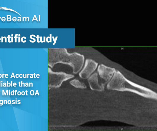
Researchers concluded that there was a significant discrepancy observed in the diagnostic abilities of the two imagingmodalities, even when readings were completed by a MSK radiologist. In the TMT joints, 15 cases were downgraded in severity, the largest discrepancy among the three groups.
Early Days: X-ray Discovery and Film-Based Imaging: The journey begins with Wilhelm Conrad Roentgen’s groundbreaking discovery of X-rays in 1895. Radiography soon became a vital tool in medicine, and for decades, it relied on photographic film to capture and develop X-ray images.
Section 1: The Shifting Paradigm in Trauma Imaging: Introduce the changing dynamics in trauma radiology, highlighting the transition from traditional imaging approaches to the emergence of extraneous imagingmodalities.
Traditional film-based X-rays gave way to digital radiography (DR) and computedradiography (CR). These digital technologies offered numerous advantages, including faster image acquisition, improved image quality, and seamless integration with electronic health records (EHRs).
Treatment Planning Because hallux valgus is a three-dimensional deformity, a three-dimensional, weight bearing imagingmodality can assist in planning surgical indications and correction.Surgeons can better determine where along the medial column the deformity should be addressed. Journal of the Foot & Ankle. 10.30795/jfootankle.2022.v16.1674.
We organize all of the trending information in your field so you don't have to. Join 5,000 users and stay up to date on the latest articles your peers are reading.
You know about us, now we want to get to know you!
Let's personalize your content
Let's get even more personalized
We recognize your account from another site in our network, please click 'Send Email' below to continue with verifying your account and setting a password.


Let's personalize your content