This site uses cookies to improve your experience. To help us insure we adhere to various privacy regulations, please select your country/region of residence. If you do not select a country, we will assume you are from the United States. Select your Cookie Settings or view our Privacy Policy and Terms of Use.
Cookie Settings
Cookies and similar technologies are used on this website for proper function of the website, for tracking performance analytics and for marketing purposes. We and some of our third-party providers may use cookie data for various purposes. Please review the cookie settings below and choose your preference.
Used for the proper function of the website
Used for monitoring website traffic and interactions
Cookie Settings
Cookies and similar technologies are used on this website for proper function of the website, for tracking performance analytics and for marketing purposes. We and some of our third-party providers may use cookie data for various purposes. Please review the cookie settings below and choose your preference.
Strictly Necessary: Used for the proper function of the website
Performance/Analytics: Used for monitoring website traffic and interactions
This is in part due to the increased messaging around how smoking is harmful to your health as well as advancements in imaging practices. Today, Low Dose ComputedTomography (LDCT) imaging is the gold standard for detecting abnormalities in the lungs because it exposes patients to less radiation than a traditional CT scan.
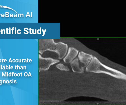
Key Points: When evaluating midfoot arthritis osteoarthritis (OA), Weight Bearing X-Ray shows many false negatives and false positives, even when read by a musculoskeletal (MSK) radiologist, as compared to weight bearing CT (WBCT). Researchers used internal data from a cohort of 302 patient feet.
Mitigating radiology workforce strain requires revisioning the specialty and adding new roles to manage imaging demand, according to a presentation delivered September 12 at the International Society for ComputedTomography (ISCT) 2024 meeting in Boston. The other role could be that of a limited x-ray machine operator.
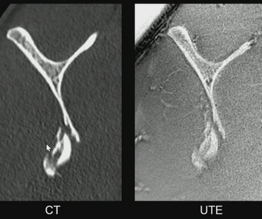
"MRI is underutilized in trauma imaging and often falls short of its potential," said presenter Prof. Sagittal view of the scapula showing a comminuted fracture of the inferior portion, imaged with computedtomography (CT, left image) and by an ultrashort time of echo (UTE) sequence (right image) on MRI.
Key Points: Cone Beam CT (CBCT) is superior in assessing bony structures compared to magnetic resonance imaging (MRI) In this study, there was a 40% rate of discrepancy when grading knee subchondral insufficiency fractures on CBCT vs. MRI, with MRI frequently underestimating damage of the subchondral bone plate while overestimating lesion size.
Teleradiology Introduction: X-ray technology has been a cornerstone of modern medicine for over a century. This blog explores the evolution, significance, and the latest advancements in X-ray technology, shedding light on how it continues to shape and revolutionize the healthcare industry.
Introduction: The history of X-rayimaging is a testament to the unceasing march of technology in healthcare. From the days of photographic film to the digital age, this blog traces the remarkable evolution of X-rayimaging, shedding light on how technology has transformed the practice of medicine.
Bracco Imaging S.p.A., part of the Bracco Group, is a global diagnostic imaging provider, headquartered in Milan, Italy, which develops, manufactures and markets diagnostic imaging agents and solutions.
Medical imaging is a crucial tool in modern healthcare, providing detailed visuals of the human body’s internal structures and helping in the accurate diagnosis and treatment of various conditions. Magnetic Resonance Imaging (MRI) MRI stands for Magnetic Resonance Imaging. X-rays are fast, painless, and commonly used.
Teleradiology & Radiology data for artificial intelligence (AI) Introduction: Embark on a journey into the world of medical imaging as we unravel the distinctions between two powerful diagnostic tools—ComputedTomography (CT) scans and Positron Emission Tomography (PET) scans.
It all started when Wilhelm Conrad Röntgen discovered X-rays in 1895. After working for weeks in his lab experimenting on the production of ‘strange rays’, which he referred to as ‘X’, he asked his wife Anna Bertha to lend ‘a hand’, the left one to be precise, which he used to produce the first X-rayimage.
Introduction of X-ray pneumoencephalography in 1947 vastly improved surgeons’ ability to localize targets, particularly with the later development of detailed stereotactic atlases. The CT and MRI images are post-contrast images and detailed planning is done to use a trajectory, which does not go through the ventricles, blood vessels.
This standard has revolutionized the radiology industry, encompassing many imagingmodalities such as X-rays, computedtomography (CT), magnetic resonance imaging (MRI), ultrasound, nuclear medicine, PET scans, etc.
Key Points: Imagingmodalities such as plain radiographs (X-Ray), computedtomography (CT), and magnetic resonance imaging (MRI), dont have the diagnostic accuracy needed to detect syndesmotic widening or subtle instability.
We organize all of the trending information in your field so you don't have to. Join 5,000 users and stay up to date on the latest articles your peers are reading.
You know about us, now we want to get to know you!
Let's personalize your content
Let's get even more personalized
We recognize your account from another site in our network, please click 'Send Email' below to continue with verifying your account and setting a password.


Let's personalize your content