This site uses cookies to improve your experience. To help us insure we adhere to various privacy regulations, please select your country/region of residence. If you do not select a country, we will assume you are from the United States. Select your Cookie Settings or view our Privacy Policy and Terms of Use.
Cookie Settings
Cookies and similar technologies are used on this website for proper function of the website, for tracking performance analytics and for marketing purposes. We and some of our third-party providers may use cookie data for various purposes. Please review the cookie settings below and choose your preference.
Used for the proper function of the website
Used for monitoring website traffic and interactions
Cookie Settings
Cookies and similar technologies are used on this website for proper function of the website, for tracking performance analytics and for marketing purposes. We and some of our third-party providers may use cookie data for various purposes. Please review the cookie settings below and choose your preference.
Strictly Necessary: Used for the proper function of the website
Performance/Analytics: Used for monitoring website traffic and interactions
His lab team is currently focused on "implementing advanced techniques, such as parallel transmit, to achieve more consistent image quality and fully harness the power of 7T for every patient," Middlebrooks said. Avoiding certain sequences due to difficulties at 7T is not always practical.
VIENNA -- It's definitely possible to develop a successful cardiac imaging practice, according to a professional development presentation delivered February 28 at the ECR in Vienna. Gutberlet described how he came to cardiac imaging early in his medical career. years of follow-up compared with CT imaging (2.1% But he persevered.
When reviewing radiographs, computedtomography (CT) scans or magnetic resonance imaging (MRI) scans, do you still turn to mnemonics every now and then to jog your short-term memory?
AI also continues to demonstrate growing utility in breast imaging, and we’ll provide coverage of key sessions in our upcoming Women’s Imaging section of the Road to RSNA. See below for previews of AI-related scientific talks we’re highlighting at this year's RSNA meeting. 10:50 a.m. | 10:30 a.m. |
Mitigating radiology workforce strain requires revisioning the specialty and adding new roles to manage imaging demand, according to a presentation delivered September 12 at the International Society for ComputedTomography (ISCT) 2024 meeting in Boston. The other role could be that of a limited x-ray machine operator.
Detecting CSIs in a clinical setting often requires imaging such as X-rays and computedtomography (CT) scans, both of which expose children to radiation, which can cause other health issues over time.
7-tesla MRI gets us closer in imaging to our pathology colleagues, and offers many new possibilities for better treatment," he said. Going forward, Middlebrooks plans to apply his skills to the further development of AI for image reconstruction. There's a huge role for AI in image acquisition," he said. Perry Pickhardt, MD.
milla1cf Fri, 02/23/2024 - 10:22 February 23, 2024 — The American Society of Radiologic Technologists (ASRT) launched its "Be Seen" campaign today to raise public awareness about the crucial role medical imaging and radiation therapy professionals play in patient diagnosis, intervention and treatment.
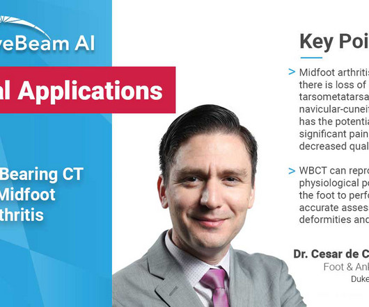
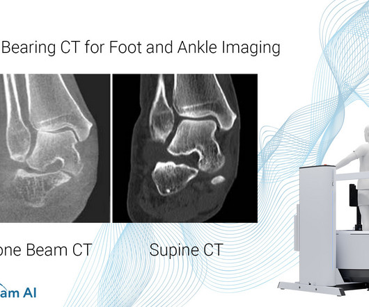
In addition, WBCT better quantifies 3 the structural deformity of Chopart, talonavicular, and calcaneocuboid joints when compared to conventional radiography and non-weight bearing computedtomographyimages. Images demonstrated severe degeneration of the midfoot joints. PMID: 33504217. (4) Iowa Orthop J. 5) Ortolani, M.,
Injecting or drinking the media contrast helps doctors see blood vessels and organs more clearly in an x-ray or a computedtomography ( CT ) scan. Qureshi said these findings highlight the need to reduce the reliance on radiographic media contrast imaging without compromising patient outcomes.
milla1cf Thu, 11/23/2023 - 06:00 November 23, 2023 — Fujifilm Healthcare Americas Corporation, a leading provider of diagnostic and enterprise imaging solutions, is unveiling several new medical systems at the 2023 Radiological Society of North America ( RSNA ) annual meeting, booth #1929, held November 26 – 30 at McCormick Place in Chicago.
1 The new FDA approved indication aligns with the recent increased focus on supplemental imaging needs for women at a higher risk for breast cancer , which may include the 40-50% of U.S. In 2019, the MEDRAD Stellant FLEX ComputedTomography ( CT ) Injection System with Certegra Workstation was also cleared in the U.S.
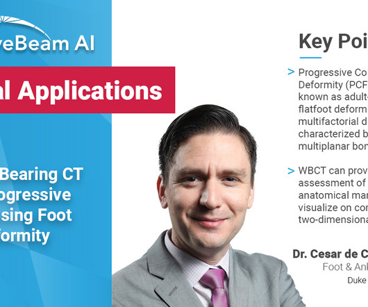
A weight bearing CT scan can: Provide an assessment of important anatomical markers of pronounced hindfoot deformity and peritalar subluxation (PTS), difficult to visualize on conventional two-dimensional radiographs 1. Weightbearing ComputedTomography for Assessment of Foot and Ankle Deformities: The Iowa Experience.
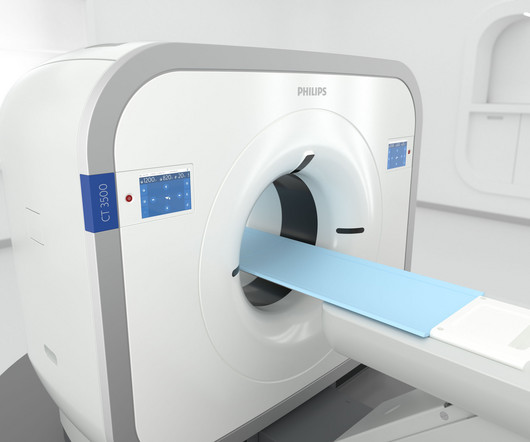
We’ve engineered the Philips CT 3500 to reduce the pain points that these high-volume departments face by developing a versatile, reliable, high-throughput imaging solution. It automates radiographers’ most time-consuming steps so that they can spend more time focusing on the patient.” Results in other cases may vary. [2]
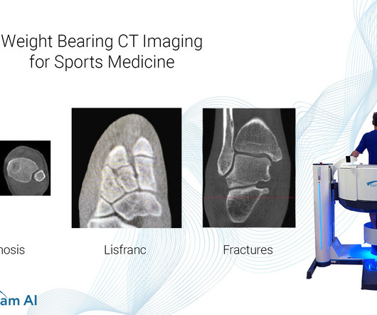
Common Indications Syndesmosis Provide increased sensitivity and specificity over radiographs 1. Flat Foot Provide an assessment of important anatomical markers of pronounced hindfoot deformity and peritalar subluxation (PTS), difficult to visualize on conventional two-dimensional radiographs 2. Foot Ankle Clin. 2023 Jun;28(2):283-295.
Common Indications Syndesmosis Provide increased sensitivity and specificity over radiographs 1. Can Weight-Bearing ComputedTomography Be a Game-Changer in the Assessment of Ankle Sprain and Ankle Instability? Insights Imaging. Help detect subtle syndesmosis injuries 1. Foot Ankle Clin. 2023 Jun;28(2):283-295.
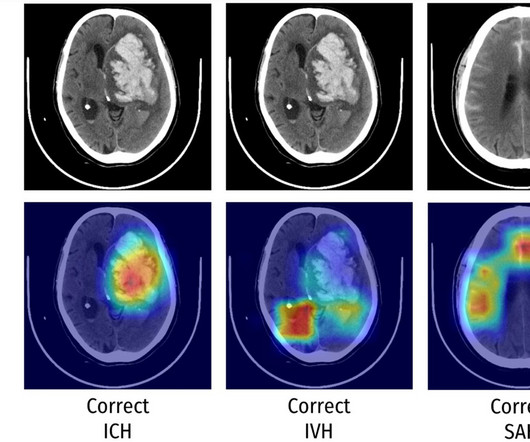
This study evaluates deep learning (DL) algorithms that are playing an increasingly important role in automatic medical image analysis. Key points The deep learning model detected intracranial haemorrhages on computedtomography with high accuracy. Chandra, Mark Brooks, Christen D. Barras & Hamed Asadi
A patient’s specific needs and concerns are assessed, and a personalized radiographic plan is developed, taking into account factors like age, health conditions, and pregnancy. Innovative Imaging Techniques: Dental radiography encompasses various imaging techniques, including intraoral, extraoral, and three-dimensional (3D) imaging.
A) AP radiograph of Lisfranc Fracture Dislocation demonstrates the circled “fleck sign” or Lisfranc ligament avulsion fracture fragment. (B) C) The lateral radiograph notes with a circle, the dorsal sub dislocation of the metatarsal base. Radiographs should be repeated after two weeks to ensure surgery is unnecessary.
Introduction: Dental X-ray technologists, also known as dental radiographers, are the unsung heroes behind the scenes of every successful dental diagnosis and treatment. Their role is critical in obtaining accurate and high-quality images of a patient’s oral and maxillofacial region.
Yes, there is an acute shortage of manpower in this space and if recent reports are anything to go by, the volume of radiographic procedures being conducted will surpass the number of radiologist being inducted into the field by a ratio of 15:2. A patient in his mid-40s had the worst headache of his life. It was around 11.30
A) Dorsoplantar radiograph of the foot demonstrating an isolated fracture of the cuboid with possible extension into the tarsometatarsal joint. (B) B) Medial oblique radiograph of the foot demonstrating an isolated fracture of the cuboid. Radiographic evidence can support the diagnosis. Trauma in a 8 year old female.
Diagnosis While radiographs are typically sufficient to make the diagnosis, WBCT scans may be useful to plan surgical treatment. Accurately assess sesamoid position as plain radiographs cannot determine whether the sesamoids have been reduced within their grooves 5. Accurately assess healing in the 1st TMT joint 4. 10.30795/jfootankle.2022.v16.1674.
What are the important findings in each image. A: Coronal CT image demonstrates normal contour of the right globe (green arrow) and a shrunken left globe (orange arrow), which is suggestive of globe rupture. C: Sagittal CT image demonstrates normal contour in the right globe (green arrow). Imaging of orbital trauma.
A: Sagittal CT image demonstrates the right posterior lens subluxation with the inferior portion of the lens displaced posteriorly into the vitreous humor (red arrow). B: Sagittal CT image demonstrates the normal location of the left lens in the iris (green arrow). Imaging of orbital trauma. Radiographics.
Additionally, the organization reported it launched its ASRT “Be Seen” public awareness campaign in late February to raise awareness about the crucial role medical imaging and radiation therapy professionals play in patient diagnosis, intervention and treatment. ASRT 2024-2025 Board of Directors Daniel DeMaio, M.Ed., Marissa Mangrum, M.S.R.S.,
Frontal abdomen radiograph demonstrates foreign body consistent with capsule endoscopy device (pill cam) in descending colon. 1 ] This diagnostic procedure involves swallowing a pill-sized camera that records thousands of images of the alimentary canal including the small intestine, an area difficult to examine via traditional endoscopy.
Applying this approach to each image cut individually can help reveal subtle findings that would otherwise be easily missed by quick scrolling. Often with parenchymal bleeds additional imaging, vascular and MRI, as well as follow up imaging will be necessary to determine the underlying cause. ComputedTomography.
Urinalysis may show pyuria, leukocytosis, nitrites, hematuria, WBC casts; however, imaging is required to confirm the diagnosis [2,3,4]. KUB indicates kidneys, ureter, and bladder (plain abdominal radiograph); CT, computedtomography; and PCD, percutaneous catheter drainage. RadioGraphics. 2000;160(6):797-805.
After working for weeks in his lab experimenting on the production of ‘strange rays’, which he referred to as ‘X’, he asked his wife Anna Bertha to lend ‘a hand’, the left one to be precise, which he used to produce the first X-ray image. after seeing the image. (2) Photoprint from radiograph by W.K. Röntgen, 1895.
Computer aided diagnostic systems are already in use throughout radiology and can accurately diagnose breast cancer with a higher degree of accuracy than their human counterparts (2) and this use will only increase. It is likely that they will lead clinic with patients to discuss their imaging, as already happens in some hospitals.
It has well-defined radiographic features and various clinical presentations. They described six radiographic phenotypes of CSVD: (1) recent small subcortical infarct, (2) white matter hyperintensity, (3) lacune of presumed vascular origin, (4) widened perivascular spaces, (5) cerebral microbleed, and (6) brain atrophy.
Key Points: Imaging modalities such as plain radiographs (X-Ray), computedtomography (CT), and magnetic resonance imaging (MRI), dont have the diagnostic accuracy needed to detect syndesmotic widening or subtle instability.
However, despite frequent use, many emergency physicians are not familiar enough with stroke imaging to interpret images on their own. Acute stroke imaging is obtained in the emergency department for two purposes. 4] These findings are consistent with other studies and highlights the limitations of NCCT in acute stroke imaging.
We organize all of the trending information in your field so you don't have to. Join 5,000 users and stay up to date on the latest articles your peers are reading.
You know about us, now we want to get to know you!
Let's personalize your content
Let's get even more personalized
We recognize your account from another site in our network, please click 'Send Email' below to continue with verifying your account and setting a password.


Let's personalize your content