This site uses cookies to improve your experience. To help us insure we adhere to various privacy regulations, please select your country/region of residence. If you do not select a country, we will assume you are from the United States. Select your Cookie Settings or view our Privacy Policy and Terms of Use.
Cookie Settings
Cookies and similar technologies are used on this website for proper function of the website, for tracking performance analytics and for marketing purposes. We and some of our third-party providers may use cookie data for various purposes. Please review the cookie settings below and choose your preference.
Used for the proper function of the website
Used for monitoring website traffic and interactions
Cookie Settings
Cookies and similar technologies are used on this website for proper function of the website, for tracking performance analytics and for marketing purposes. We and some of our third-party providers may use cookie data for various purposes. Please review the cookie settings below and choose your preference.
Strictly Necessary: Used for the proper function of the website
Performance/Analytics: Used for monitoring website traffic and interactions
ASRT's survey confirmed that all major disciplines in medical imaging and radiation therapy in the U.S. Magnetic resonance imaging technologists saw an increase of 12.5% Computedtomography and radiography each experienced a 12.3% have seen wage and salary increases since 2022. increase since 2022.
7-tesla MRI gets us closer in imaging to our pathology colleagues, and offers many new possibilities for better treatment," he said. Going forward, Middlebrooks plans to apply his skills to the further development of AI for image reconstruction. There's a huge role for AI in image acquisition," he said. Perry Pickhardt, MD.
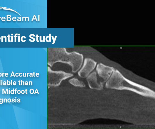
Researchers concluded that there was a significant discrepancy observed in the diagnostic abilities of the two imaging modalities, even when readings were completed by a MSK radiologist. In the TMT joints, 15 cases were downgraded in severity, the largest discrepancy among the three groups.
This annual event recognizes the hard work and dedication of radiologic technologists (RTs) who operate imaging technology to aid in accurate diagnoses and treatments. For instance, radiography reported an 18.1% vacancy rate, while computedtomography had a 17.7% vacancy rate.
Diagnostic imaging is an important tool used every day in healthcare to assist doctors in making the most informed decisions for their patients. Over the years, advancements in diagnostic imaging have greatly increased patients’ overall care, quality of life, and outcome when diagnosed with certain conditions.
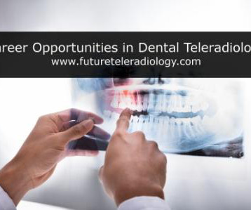
Teleradiology Introduction: Dental radiography plays a pivotal role in modern dentistry, enabling practitioners to diagnose and treat various oral health conditions. However, the field of dental radiography has undergone significant advancements, along with its own set of challenges and triumphs.
This is one of the topics to be discussed by KA Imaging’s Team Lead for Spectral Imaging, Steven Tilley , during a presentation at the upcoming European Congress of Radiology ( ECR ). The objective was to measure their performance reading Digital Radiography versus Dual-Energy images by the Reveal 35C. said CTO Karim S.
milla1cf Thu, 11/23/2023 - 06:00 November 23, 2023 — Fujifilm Healthcare Americas Corporation, a leading provider of diagnostic and enterprise imaging solutions, is unveiling several new medical systems at the 2023 Radiological Society of North America ( RSNA ) annual meeting, booth #1929, held November 26 – 30 at McCormick Place in Chicago.
This is one of the topics to be discussed by KA Imaging’s Team Lead for Spectral Imaging, Steven Tilley , during a presentation at the upcoming European Congress of Radiology ( ECR ). The objective was to measure their performance reading Digital Radiography versus Dual-Energy images by the Reveal 35C. said CTO Karim S.
milla1cf Fri, 02/23/2024 - 10:22 February 23, 2024 — The American Society of Radiologic Technologists (ASRT) launched its "Be Seen" campaign today to raise public awareness about the crucial role medical imaging and radiation therapy professionals play in patient diagnosis, intervention and treatment.
MRI-Scan-Teleradiology Introduction: Dental radiography is an essential component of modern dentistry, offering valuable insights into oral health and guiding treatments with precision. Patient-Centered Approach: Dental radiography starts with the patient. Cone-Beam ComputedTomography (CBCT): CBCT is a game-changer in dental imaging.
In addition, WBCT better quantifies 3 the structural deformity of Chopart, talonavicular, and calcaneocuboid joints when compared to conventional radiography and non-weight bearing computedtomographyimages. Images demonstrated severe degeneration of the midfoot joints. PMID: 33504217. (4) Iowa Orthop J.
Here you’ll find a highly skilled, compassionate staff along with comprehensive advanced imaging technologies that are raising the bar for patient care in their rural community. Only 21% of extremely disadvantaged zip codes had access to computedtomography (CT) facilities as compared to 32% of extremely advantaged zip codes.
Patients with metal implants can obtain advanced diagnostic imaging. Low Dose “Considering the low dose of radiation and high image quality, CBCT could be used as a priority method of choice to assess the structure of wrist and hand bones and be done as the first step in diagnostics, replacing standard radiography.
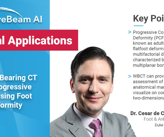
In addition, WBCT better quantifies the structural deformity of PCFD compared to conventional radiography and non-weight bearing CT images 1 . Weightbearing ComputedTomography for Assessment of Foot and Ankle Deformities: The Iowa Experience. PMID: 33504217. (4) Iowa Orthop J. 2021;41(1):111-119. 5) Ortolani, M.,
Introduction: The history of X-ray imaging is a testament to the unceasing march of technology in healthcare. From the days of photographic film to the digital age, this blog traces the remarkable evolution of X-ray imaging, shedding light on how technology has transformed the practice of medicine.
Teleradiology Introduction: “Beneath the Surface” is an illuminating journey that delves deep into the world of X-ray imaging, unveiling its crucial role in patient care. In this exploration, we will dive into the history, technology, and the profound impact of X-ray imaging on healthcare.
a Sunnyvale, CA-based developer of a next generation artificial intelligence (AI)-based tomosynthesis X-ray imaging system, has reported that its team, which began clinical trials in late 2023, is very pleased with the early ARC60 imaging results, both in terms of depiction of details and consistency of imaging quality.
Introduction: The realm of diagnostic imaging has taken a revolutionary leap with the introduction of Cone Beam ComputedTomography (CBCT). This technology has paved the way for navigating the third dimension, offering a transformative role in imaging that transcends the limitations of conventional two-dimensional radiography.
Teleradiology Introduction: The field of dentistry is undergoing a digital revolution, and at the forefront of this transformation is the realm of dental X-ray imaging. In this article, we will explore the latest innovations that are shaping the future of dental X-ray imaging, revolutionizing patient care and clinical practices.
Closeup of X-ray photography of human brain Introduction: “The Radiant Thread” is a comprehensive guide that unravels the intricate world of X-ray imaging from A to Z. From its historical roots to contemporary innovations, we will follow the radiant thread that connects all aspects of X-ray imaging.
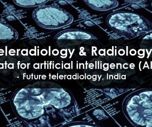
Teleradiology & Radiology data for artificial intelligence (AI) Introduction: “Illuminating Shadows” invites you on a comprehensive journey into the fascinating world of X-ray imaging. Chapter 1: Introduction to X-ray Imaging An overview of the importance of X-ray imaging in healthcare.
How X-rays are generated, interact with the human body, and create diagnostic images. Chapter 3: The Evolution of Radiography: From Shadows to Images An exploration of the development of radiography, the earliest X-ray imaging technique.
This journey takes us from the early days of X-ray discovery by Wilhelm Roentgen to the cutting-edge digital and computational innovations that shape the modern landscape of diagnostic imaging. Chapter 2: The Art and Science of Radiography A closer look at the development of radiography, the first X-ray imaging method.
From Film to Digital: The transition from film-based X-ray systems to digital technology marked a turning point in dental radiography. Digital sensors and intraoral cameras offer instant image acquisition, reduced radiation exposure, and the ability to enhance and manipulate images for better diagnostics.
MRI-Scan-Teleradiology Introduction: In “Trauma Radiology Unveiled: Examining the Emergence of Extraneous Imaging as the New Norm,” we embark on a detailed exploration of the evolving landscape of trauma radiology.
The historical backdrop of Wilhelm Roentgen’s serendipitous discovery and the birth of radiography. How X-rays are generated, interact with matter, and produce diagnostic images. Chapter 4: Inside the X-ray Machine: The Art of Image Production A closer look at the components and mechanics of X-ray machines.
Traditional film-based X-rays gave way to digital radiography (DR) and computedradiography (CR). These digital technologies offered numerous advantages, including faster image acquisition, improved image quality, and seamless integration with electronic health records (EHRs).
At GE, he gained deep expertise in ComputedTomography (CT) and Radiography systems, with a significant focus on the design and manufacturing of x-ray tubes, high-voltage generators, and x-ray detectors. The post Eric Chappelear, Vice President Facility Operations appeared first on TTG Imaging Solutions.
Discuss the initial applications of radiography and its profound impact on the landscape of medical diagnostics. Technological Metamorphosis: From Analog to Digital Prowess Chart the transition from analog radiology to the epoch of digital imaging technologies.
Introduction: While X-rays are traditionally associated with skeletal imaging, their reach extends far beyond bones. Angiography, a technique that employs contrast agents and X-rays, provides detailed images of blood vessels, enabling the diagnosis and treatment of various heart and vascular conditions.
Their role is critical in obtaining accurate and high-quality images of a patient’s oral and maxillofacial region. Image Capture: Precise Positioning: Proper positioning of the X-ray machine and the patient is crucial. The technologist ensures that the images are of high quality and can be accurately interpreted by the dentist.
Additionally, the organization reported it launched its ASRT “Be Seen” public awareness campaign in late February to raise awareness about the crucial role medical imaging and radiation therapy professionals play in patient diagnosis, intervention and treatment. ASRT 2024-2025 Board of Directors Daniel DeMaio, M.Ed., Jennifer Thompson, Ed.D.,
Medical imaging of the human skeleton enables accurate diagnosis, treatment, and monitoring of diverse bone and joint conditions. The post Medical Imaging of the Human Skeleton appeared first on Open Medscience.
A: Sagittal CT image demonstrates the right posterior lens subluxation with the inferior portion of the lens displaced posteriorly into the vitreous humor (red arrow). B: Sagittal CT image demonstrates the normal location of the left lens in the iris (green arrow). Imaging of orbital trauma. Xray of the Week Figure 1.
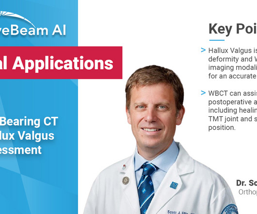
Treatment Planning Because hallux valgus is a three-dimensional deformity, a three-dimensional, weight bearing imaging modality can assist in planning surgical indications and correction.Surgeons can better determine where along the medial column the deformity should be addressed. Journal of the Foot & Ankle. 10.30795/jfootankle.2022.v16.1674.
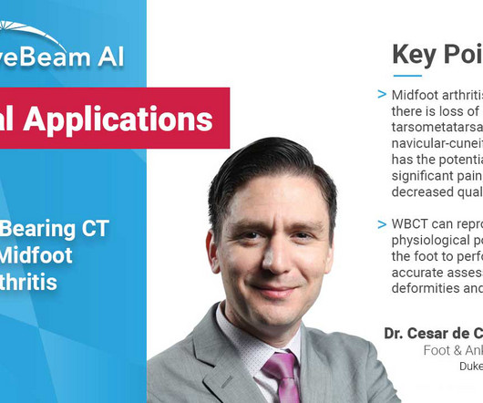
Imaging and Case Analysis: Radiographic images demonstrate misalignment of the medial side of the second metatarsal with the medial side of the middle cuneiform bone, as seen in this case. A computedtomography scan will better assist with diagnosis and help with planning if surgery is necessary [3]. J Foot Ankle Res.
1] Using a well validated decision instrument, such as the Canadian CT Head Rule in adults or the PECARN rule in children, reduces the frequency of unnecessary imaging and decreases length of stay while increasing the diagnostic yield (frequency of positive tests) amongst those patients that are imaged.[2,3] 2005 Oct;59(4):954-9.
We organize all of the trending information in your field so you don't have to. Join 5,000 users and stay up to date on the latest articles your peers are reading.
You know about us, now we want to get to know you!
Let's personalize your content
Let's get even more personalized
We recognize your account from another site in our network, please click 'Send Email' below to continue with verifying your account and setting a password.


Let's personalize your content