This site uses cookies to improve your experience. To help us insure we adhere to various privacy regulations, please select your country/region of residence. If you do not select a country, we will assume you are from the United States. Select your Cookie Settings or view our Privacy Policy and Terms of Use.
Cookie Settings
Cookies and similar technologies are used on this website for proper function of the website, for tracking performance analytics and for marketing purposes. We and some of our third-party providers may use cookie data for various purposes. Please review the cookie settings below and choose your preference.
Used for the proper function of the website
Used for monitoring website traffic and interactions
Cookie Settings
Cookies and similar technologies are used on this website for proper function of the website, for tracking performance analytics and for marketing purposes. We and some of our third-party providers may use cookie data for various purposes. Please review the cookie settings below and choose your preference.
Strictly Necessary: Used for the proper function of the website
Performance/Analytics: Used for monitoring website traffic and interactions
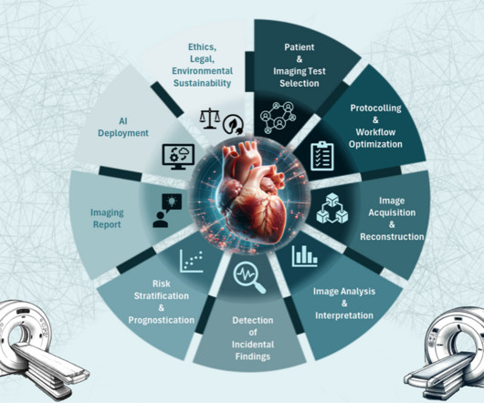
To better assess the current landscape and challenges of AI applications in cardiac CT and MRI specifically, the paper aims to bridge the gap between burgeoning research developments and limited clinical applications, according to lead author Domenico Mastrodicasa, MD, from the University of Washington in Seattle, and colleagues.
Middlebrooks' research interest consists of using ultrahigh-field, 7-tesla MRI to plot brain microstructure and develop surgical treatment of brain tumors, epilepsy, and neurodegenerative and movement disorders such as Parkinson's disease, essential tremor, and dystonia. Middlebrooks, MD, of the Mayo Clinic in Jacksonville, FL.
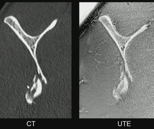
SINGAPORE -- The use of MRI for musculoskeletal (MSK) trauma imaging is becoming increasingly viable, according to a presentation delivered May 7 at the International Society of Magnetic Resonance in Imaging (ISMRM) meeting. MRI is underutilized in trauma imaging and often falls short of its potential," said presenter Prof.
Magnetic Resonance Imaging (MRI) This type of diagnostic imaging, commonly known as MRI , uses powerful magnetic fields and radio waves to create detailed images of the internal body. Unlike MRIs, an X-Ray only requires a few minutes before the results are examined by a radiologist and sent to your primary care doctor.
. | S2-SSGI01-2 | E451B Transparently benchmarked, expert AI can enable opportunistic screening to start catching pancreatic ductal adenocarcinoma (PDAC) on contrast-enhanced computedtomography (CECT) at earlier stages, according to first results of the PANORAMA (Pancreatic cancer diagnosis: Radiologists meet AI) study. 11:00 a.m. |
At the time, there was little training for, say, cardiac MRI. cardiac CT or MRI, nuclear cardiology, or echocardiology) for myocardial ischemia and the more invasive coronary CT angiography (CTA) for patients exhibiting symptoms of the disease and for whom obstructive CAD can't be excluded by clinical assessment alone.
discussed a variety of imaging features associated with cerebrospinal fluid (CSF) leaks, the diagnostic utility of the Bern score as well as the merits of 3D T2FS MRI and dynamic computedtomography myelography. In a recent lecture at the American Roentgen Ray Society (ARRS) 2023 Annual Meeting, Andrew Callen, M.D.,
When reviewing radiographs, computedtomography (CT) scans or magnetic resonance imaging (MRI) scans, do you still turn to mnemonics every now and then to jog your short-term memory?
Computedtomography (CT scans) plays a critical role in diagnosing and monitoring a wide range of medical conditions. MRI Isn’t An Option Another form of diagnostic imaging is Magnetic Resonance Imaging (MRI) , which is used to diagnose brain and spinal cord disorders, joint and musculoskeletal conditions, and cancer.
Key Points: Cone Beam CT (CBCT) is superior in assessing bony structures compared to magnetic resonance imaging (MRI) In this study, there was a 40% rate of discrepancy when grading knee subchondral insufficiency fractures on CBCT vs. MRI, with MRI frequently underestimating damage of the subchondral bone plate while overestimating lesion size.
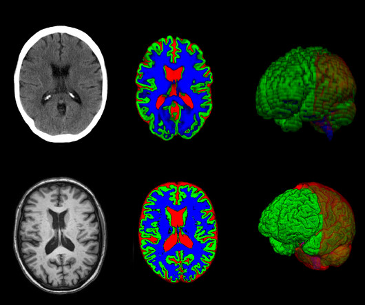
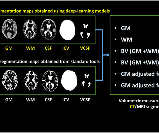
milla1cf Fri, 10/20/2023 - 18:29 October 20, 2023 — In certain cases, a new method can provide as much information from brain images taken with computedtomography (CT) as images captured with magnetic resonance imaging (MRI). It has been created using deep learning, a form of artificial intelligence (AI).
In certain cases, a new method can provide as much information from brain images taken with computedtomography (CT) as images captured with magnetic resonance imaging (MRI). The method could enhance diagnostic support, particularly in primary care, for conditions such as dementia and other brain disorders.
His research interests include using structural and functional MRI -- particularly ultrahigh-field, 7-tesla MRI -- to map brain microstructure and develop neurosurgical treatment of brain tumors, epilepsy, and neurodegenerative and movement disorders such as Parkinson's disease, essential tremor, and dystonia.
In certain cases, a new method can provide as much information from brain images taken with computedtomography as images captured with magnetic resonance imaging.
The Radiological Society of North America’s (RSNA) 2024 annual meeting showcased significant advancements in medical imaging, including artificial intelligence (AI), innovations in computedtomography (CT) and magnetic resonance imaging (MRI), and strategies to address the ongoing radiology staffing shortage.
discussed key challenges with the use of SPECT MRI and how an emerging deep learning model may facilitate attenuation compensation without the need for an additional computedtomography (CT) scan. In a recent interview, Abhinav K.
Photon-counting CT (PCCT) technology has the potential to revolutionize cardiac imaging, according to a presentation delivered July 20 at the Society for Cardiovascular ComputedTomography (SCCT) annual meeting in Washington, DC. Next-generation cardiac CT will become more like MRI. "We
If its been a few years since your team has opted for a new ComputedTomography (CT) scanner, you might be surprised by the progress the technology has made.
Your healthcare facility has decided to invest in a new computedtomography (CT) scanner; now you’re in the process of evaluating your options. While considering variables like imaging capabilities, workflow efficiency, and patient experience, you’re also crunching numbers tied to cost.
SPE is not well visualized with current non-invasive imaging tools (ultrasound and MRI) and definitive diagnosis requires laparoscopy. tim.hodson Wed, 02/05/2025 - 09:56 Feb. 5, 2025 Serac Healthcare Ltd., Initial Phase II findings indicate that 99m Tc-maraciclatide has potential as a non-invasive test for the detection of SPE.
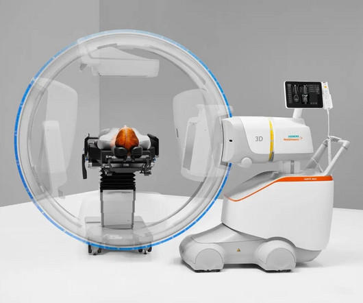
The system accelerates and standardizes 2D fluoroscopic and 3D cone-beam computedtomography (CT) imaging for surgeons and operating room teams in hospitals and outpatient facilities, bringing consistency to automated workflows and reducing imaging time during operations.
Computedtomography (CT scans) plays a critical role in diagnosing and monitoring a wide range of medical conditions. MRI Isn’t An Option Another form of diagnostic imaging is Magnetic Resonance Imaging (MRI) , which is used to diagnose brain and spinal cord disorders, joint and musculoskeletal conditions, and cancer.
broken bones) Utilizing diagnostic imaging techniques such as digital radiography, computedtomography (CT) scans , endoscopy, ultrasound, and magnetic resonance imaging (MRI) can help monitor disease progression as well as aid in early detection by non-invasive or minimally invasive techniques that require little to no recovery time.
The long-anticipated advancement is made achievable through the adaptation of tumor imaging processes employing positron computedtomography. A more refined approach to the precise removal of brain tumors is on the horizon.
milla1cf Thu, 01/04/2024 - 10:47 January 4, 2024 — Diagnosing cancer today involves using chemical “contrast agents” to improve the accuracy of medical imaging processes such as X-rays as well as computedtomography (CT) and magnetic resonance imaging (MRI) scans.
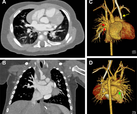
A comprehensive assessment, including ultrasound, MRI and CT exams, is typically needed to plan for surgery and to create virtual and printed 3D reconstructions of the heart. Congenital heart defects are the leading cause of morbidity and mortality in the neonatal period, occurring in up to one percent of live births.
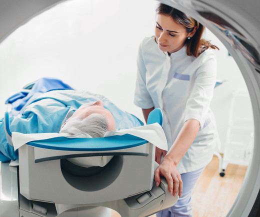
MRI Scan An MRI scan is a noninvasive, safe way for healthcare professionals to examine a patient's organs, tissues, and skeletal system. Radio waves cause these realigned atoms to produce signals, which are used to create the cross-sectional MRI images. The answer will depend upon the type of MRI being performed.
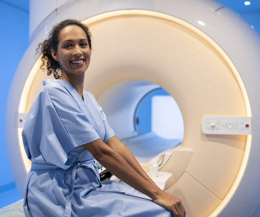
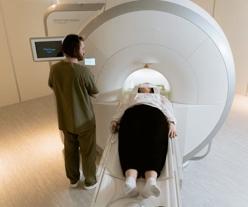
Magnetic resonance imaging (MRI) is used to help diagnose and treat various medical conditions. At Intermountain Medical Imaging , we rely on a variety of MRI options that offer a wider opening, helping us deliver high-quality care to clients and medical providers alike. What Is a Wide Bore MRI?
Techniques such as mammography, low-dose computedtomography (LDCT), and magnetic resonance imaging (MRI) are instrumental in identifying cancers like breast, lung, and prostate in their nascent stages.
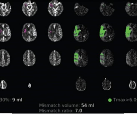
Unvaccinated people with COVID-19 who undergo angiographic reperfusion after acute ischemic stroke may have a greater than fivefold risk of continued infarct growth in comparison to unvaccinated people without COVID-19, according to computedtomography perfusion (CTP) and magnetic resonance imaging (MRI) findings from a recently published study.
Bracco Imaging S.p.A., part of the Bracco Group, is a global diagnostic imaging provider, headquartered in Milan, Italy, which develops, manufactures and markets diagnostic imaging agents and solutions.
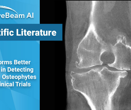
Key Points: While magnetic resonance imaging (MRI) adequately detects the size and presence of osteophytes (OPs) in the medial tibio-femoral compartment, it underestimates OPs in all other knee compartments of osteoarthritic patients. Clinical trials will often use MRI to assess and grade OPs in arthritic knees. Roemer et al.
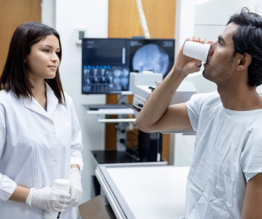
Injecting or drinking the media contrast helps doctors see blood vessels and organs more clearly in an x-ray or a computedtomography ( CT ) scan. Some suggested methods include doing more magnetic resonance imaging ( MRI ) scans and reducing the amount of dye used per patient.
The approval expands upon Bayer's focus on breast imaging, with a portfolio that also includes Gadavist (gadobutrol) injection, a gadolinium-based contrast agent approved for use with MRI ( Magnetic Resonance Imaging ) to assess the presence and extent of malignant breast disease in adult patients. for use in CEM.
2 TFCC Tear Patient injected with 1cc or less of contrast, as opposed to 8-10cc required for an MRI exam. The use of cone-beam computedtomography (CBCT) in radiocarpal fractures: a diagnostic test accuracy meta-analysis. Capabilities of Cone-Beam ComputedTomography in the Assessment of the Structure of Wrist and Hand Bones.
In this blog post, we’ll explore the differences and uses of MRI, CT scans, X-rays, ultrasounds, and PET/CT to help you better understand what to expect and how these technologies can assist in your healthcare. Magnetic Resonance Imaging (MRI) MRI stands for Magnetic Resonance Imaging.
Advanced scanner with ultra-fast time of flight and unique air-cooled design Sustainable imaging platform with low total cost of ownership Addresses all clinical needs, including oncology, theranostics, cardiology, and neurology Siemens Healthineers announces the Food and Drug Administration clearance of the Biograph Trinion, a high-performance, energy-efficient (..)
WBCT scans can also be used as an adjunct to an ankle MRI to correlate anatomic abnormalities with functional instability. Assess for instability, especially in cases with accompanying Weber B fractures 2 . Diagnosis If WB X-rays are indeterminate after clinical exam, order a bilateral WBCT.
MRI system and a 128- slice computedtomography (CT) system. “At Next generation MRI with the APERTO Lucent Plus*: MR imaging technology from APERTO Lucent Plus is based on a permanent magnet system providing a 0.4T less than systems in its class.
™¹, a positron emission tomography/computedtomography (PET/CT) scanner with a time of flight (TOF) of 178 picoseconds (ps)—the industry’s fastest TOF.² The next-generation system builds on the established performance of the Biograph Vision family of scanners.
We organize all of the trending information in your field so you don't have to. Join 5,000 users and stay up to date on the latest articles your peers are reading.
You know about us, now we want to get to know you!
Let's personalize your content
Let's get even more personalized
We recognize your account from another site in our network, please click 'Send Email' below to continue with verifying your account and setting a password.


Let's personalize your content