This site uses cookies to improve your experience. To help us insure we adhere to various privacy regulations, please select your country/region of residence. If you do not select a country, we will assume you are from the United States. Select your Cookie Settings or view our Privacy Policy and Terms of Use.
Cookie Settings
Cookies and similar technologies are used on this website for proper function of the website, for tracking performance analytics and for marketing purposes. We and some of our third-party providers may use cookie data for various purposes. Please review the cookie settings below and choose your preference.
Used for the proper function of the website
Used for monitoring website traffic and interactions
Cookie Settings
Cookies and similar technologies are used on this website for proper function of the website, for tracking performance analytics and for marketing purposes. We and some of our third-party providers may use cookie data for various purposes. Please review the cookie settings below and choose your preference.
Strictly Necessary: Used for the proper function of the website
Performance/Analytics: Used for monitoring website traffic and interactions
Middlebrooks' research interest consists of using ultrahigh-field, 7-tesla MRI to plot brain microstructure and develop surgical treatment of brain tumors, epilepsy, and neurodegenerative and movement disorders such as Parkinson's disease, essential tremor, and dystonia. I'm a radiographer,' " Stewart recalled.
At the time, there was little training for, say, cardiac MRI. Together with our radiographers, I learned to scan cardiac patients and learned special anatomy from pediatric cardiologists and pediatric cardiac surgeons." Gutberlet described how he came to cardiac imaging early in his medical career. But he persevered.
When reviewing radiographs, computedtomography (CT) scans or magnetic resonance imaging (MRI) scans, do you still turn to mnemonics every now and then to jog your short-term memory?
. | S1-SSCH01-5 | E451A This scientific paper may increase overall confidence in the potential of using multimodal AI for tuberculosis (TB) detection, and potentially autonomous reporting, on chest radiographs in certain clinical settings. 3D body composition analysis of whole-body MRI can predict mortality Wednesday, December 4 | 10:06 a.m.-10:18
His research interests include using structural and functional MRI -- particularly ultrahigh-field, 7-tesla MRI -- to map brain microstructure and develop neurosurgical treatment of brain tumors, epilepsy, and neurodegenerative and movement disorders such as Parkinson's disease, essential tremor, and dystonia.
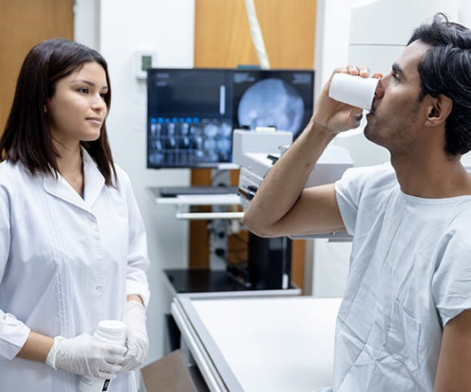
Injecting or drinking the media contrast helps doctors see blood vessels and organs more clearly in an x-ray or a computedtomography ( CT ) scan. Qureshi said these findings highlight the need to reduce the reliance on radiographic media contrast imaging without compromising patient outcomes.
The approval expands upon Bayer's focus on breast imaging, with a portfolio that also includes Gadavist (gadobutrol) injection, a gadolinium-based contrast agent approved for use with MRI ( Magnetic Resonance Imaging ) to assess the presence and extent of malignant breast disease in adult patients. RadioGraphics 2019 39:7, 1907-1920.
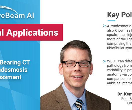
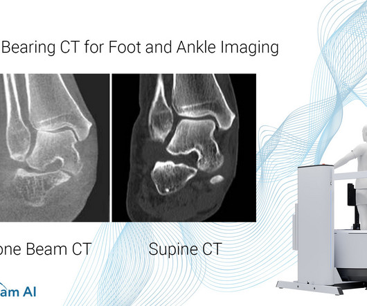
A weight bearing CT scan can: Provide increased sensitivity and specificity over radiographs. WBCT scans can also be used as an adjunct to an ankle MRI to correlate anatomic abnormalities with functional instability. Diagnosis If WB X-rays are indeterminate after clinical exam, order a bilateral WBCT.
MRI system and a 128- slice computedtomography (CT) system. “At The Essentia SA is an ultra-compact straight arm system, designed for a wide range of standing, sitting and recumbent radiographic exams. less than systems in its class. static field strength and a space-saving, patient-comfortable gantry design.
Common Indications Syndesmosis Provide increased sensitivity and specificity over radiographs 1. Flat Foot Provide an assessment of important anatomical markers of pronounced hindfoot deformity and peritalar subluxation (PTS), difficult to visualize on conventional two-dimensional radiographs 2. Foot Ankle Clin. 2023 Jun;28(2):283-295.
MRI-Scan-Teleradiology Introduction: Dental radiography is an essential component of modern dentistry, offering valuable insights into oral health and guiding treatments with precision. Cone-Beam ComputedTomography (CBCT): CBCT is a game-changer in dental imaging. Let’s explore the key aspects of this intricate process.
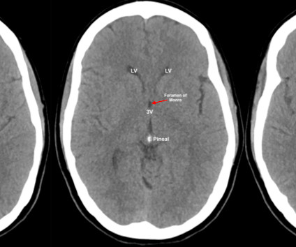
Yes, there is an acute shortage of manpower in this space and if recent reports are anything to go by, the volume of radiographic procedures being conducted will surpass the number of radiologist being inducted into the field by a ratio of 15:2. A patient in his mid-40s had the worst headache of his life. It was around 11.30
The goal of this multi-part blog is as follows: To cover the basics of how to look at a CT brain and quickly identify life threat Review the literature supporting the major ED indications Discuss special considerations, such as when to use contrast, angiography, or MRI instead. ComputedTomography. Shaw AS, Prokop M. McKetty MH.
Photoprint from radiograph by W.K. 3) In the early twentieth century, it was a common goal for investigators to try to find a way to separate the superimposed shadows that were recorded when a complex structure was shown on a radiograph. (3) 3) This is what is known as tomography. This is now known as ‘Hand mit Ringen’. (1)
A trial, which involved analysing a computerised tomography (CT) scan of a patient’s lungs against three expert human radiologists, found that Enlitic’s system was 50% better at classifying malignant tumours and had a false-negative rate of zero, compared with 7% for the humans (3). 2011, Radiographics, pp. Hough, Andrew.
It has well-defined radiographic features and various clinical presentations. They described six radiographic phenotypes of CSVD: (1) recent small subcortical infarct, (2) white matter hyperintensity, (3) lacune of presumed vascular origin, (4) widened perivascular spaces, (5) cerebral microbleed, and (6) brain atrophy.
Key Points: Imaging modalities such as plain radiographs (X-Ray), computedtomography (CT), and magnetic resonance imaging (MRI), dont have the diagnostic accuracy needed to detect syndesmotic widening or subtle instability.
It has a similar sensitivity and specificity for acute ischemic stroke as CTA, its use has been validated in multiple interventional stroke studies, and it has been shown to predict core infarct size accurately compared to the gold standard MRI.[7,8,15] Radiographics. Approach to reperfusion therapy for acute ischemic stroke.
We organize all of the trending information in your field so you don't have to. Join 5,000 users and stay up to date on the latest articles your peers are reading.
You know about us, now we want to get to know you!
Let's personalize your content
Let's get even more personalized
We recognize your account from another site in our network, please click 'Send Email' below to continue with verifying your account and setting a password.


Let's personalize your content