This site uses cookies to improve your experience. To help us insure we adhere to various privacy regulations, please select your country/region of residence. If you do not select a country, we will assume you are from the United States. Select your Cookie Settings or view our Privacy Policy and Terms of Use.
Cookie Settings
Cookies and similar technologies are used on this website for proper function of the website, for tracking performance analytics and for marketing purposes. We and some of our third-party providers may use cookie data for various purposes. Please review the cookie settings below and choose your preference.
Used for the proper function of the website
Used for monitoring website traffic and interactions
Cookie Settings
Cookies and similar technologies are used on this website for proper function of the website, for tracking performance analytics and for marketing purposes. We and some of our third-party providers may use cookie data for various purposes. Please review the cookie settings below and choose your preference.
Strictly Necessary: Used for the proper function of the website
Performance/Analytics: Used for monitoring website traffic and interactions
Sites can choose to send CT studies directly from CT machines, with a routing rule in PACS, or with a DICOM router. International variation in radiation dose for computedtomography examinations: prospective cohort study. All calculations are performed without the data leaving the provider’s network. 2019 Jan 2:364:k4931.
Whether it’s the proverbial “bad penny” case that crashes PACS or the perception that positive computedtomography angiography (CTA) exams for pulmonary arterial clots come in sets of three, suspicions can emerge here and there in radiology.
Radiation doses from computedtomography (CT) scans on patients are highly variable across patients and hospitals throughout the United States and other nations.1 International variation in radiation dose for computedtomography examinations: prospective cohort study. BMJ (Clinical research ed). Jan 2019;364:k4931.
Spotlight key technological breakthroughs, including computedtomography (CT), magnetic resonance imaging (MRI), and ultrasound, revolutionizing the diagnostic panorama. Discuss how PACS has redefined collaboration and accessibility within the dynamic realm of radiology workflows.
Chapter 4: Beyond Radiography: Advanced X-ray Modalities An examination of advanced X-ray modalities, including fluoroscopy, computedtomography (CT), and mammography. How PACS streamlines image access, reporting, and collaboration among healthcare professionals.
The Radiology Experience Tour expanded this year to include a portfolio of Philips imaging solutions including Magnetic Resonance, Radiology and Fluoroscopy, Ultrasound, Radiology Operations Command Center (ROCC), PACS, C-Arms and Ambient Experience. The objectives behind the radiology experience tour are twofold.
Chapter 3: The Radiologic Toolbox – Types of X-ray Imaging An exploration of the various types of X-ray imaging, including radiography, fluoroscopy, computedtomography (CT), and more. How PACS streamlines image access and reporting, improving healthcare efficiency.
Chapter 5: Beyond Radiography: The Rise of Advanced Modalities An examination of advanced X-ray modalities, including fluoroscopy, mammography, and computedtomography (CT). How PACS has streamlined image access and reporting, improving healthcare efficiency.
Chapter 4: Beyond Radiography: Advanced X-ray Modalities An examination of advanced X-ray modalities, including fluoroscopy, computedtomography (CT), and mammography. How PACS streamlines image access, reporting, and collaboration among healthcare professionals.
DICOM allows transmitting medical imaging data to devices like scanners, servers, workstations, printers, network hardware, and PACS. In this blog article, we will explain the fundamentals of DICOM technology, its impact on clinical practice and radiology workflows, and dig deeper into enhanced functionality offered by modern PACS systems.
Like PACS , CPACS systems typically use DICOM (Digital Imaging and Communications in Medicine) standards to store and transmit images. Cardiac CT is a type of computedtomography used to image the heart. It is a medical imaging system used to store, manage, and share cardiology images. How Does CPACS Work?
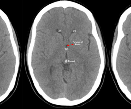
This can be done manually or, typically, PACS viewers have preset brain, bone, lung and soft tissue windows that can be displayed by pressing different numbers on the keypad. ComputedTomography. Demystifying the Hand Exam. Expert Commentary by Rusinak D ]. Retrieved from [link] Posts you may also enjoy References 1. McKetty MH.
Tomography originates from the Greek words ‘tomos’, meaning ‘slice’ or ‘section’, and ‘graphia’, meaning ‘description of’. In the 1960s computers were increasingly available and more powerful and in 1971, the first computedtomography (CT) scan was performed on a patient.
We organize all of the trending information in your field so you don't have to. Join 5,000 users and stay up to date on the latest articles your peers are reading.
You know about us, now we want to get to know you!
Let's personalize your content
Let's get even more personalized
We recognize your account from another site in our network, please click 'Send Email' below to continue with verifying your account and setting a password.


Let's personalize your content