This site uses cookies to improve your experience. To help us insure we adhere to various privacy regulations, please select your country/region of residence. If you do not select a country, we will assume you are from the United States. Select your Cookie Settings or view our Privacy Policy and Terms of Use.
Cookie Settings
Cookies and similar technologies are used on this website for proper function of the website, for tracking performance analytics and for marketing purposes. We and some of our third-party providers may use cookie data for various purposes. Please review the cookie settings below and choose your preference.
Used for the proper function of the website
Used for monitoring website traffic and interactions
Cookie Settings
Cookies and similar technologies are used on this website for proper function of the website, for tracking performance analytics and for marketing purposes. We and some of our third-party providers may use cookie data for various purposes. Please review the cookie settings below and choose your preference.
Strictly Necessary: Used for the proper function of the website
Performance/Analytics: Used for monitoring website traffic and interactions
While the American College of Radiology (ACR) publishes typical observed doses in its Dose Index Registry , there are few published doses that are defined as excessive, and without a defined passing dose, it is hard for a physician to calibrate doses. 2019 Jan 2:364:k4931. Smith-Bindman R, Chu P, Wang Y, et al. JAMA Intern Med.
It specifies the adoption of a new quality measure into the diagnostic radiology set of the Quality Payment Program (QPP) called Excessive Radiation Dose or Inadequate Image Quality for Diagnostic ComputedTomography (CT) in Adults, with the goal of improving patientsafety by optimizing CT radiation dose.
Imalogix is highlighting a January 30 update regarding the patientsafety Excessive Radiation (ExRad) Dose or Inadequate Image Quality for Diagnostic ComputedTomography in Adults electronic clinical quality measure (eCQM).
The software enables users to connect to computedtomography (CT), magnetic resonance (MR), positron emission tomography (PET), single-photon emission CT (SPECT), PET/CT , SPECT/CT, and PET/MR scanners from Siemens Healthineers as well as other equipment vendors, regardless of location.
tim.hodson Fri, 08/09/2024 - 15:45 At the annual AHRA (American Healthcare Radiology Administrators) conference in Orlando, Florida, Bayer announced an exploratory collaboration with Alara Imaging, Inc. with these important patientsafety measures," Nate Mazonson, CEO and co-founder, Alara Imaging, Inc. Jan 2019;364:k4931.
Its SunCHECK Platform standardizes workflows and centralizes data management for patient and machine quality assurance in radiation therapy, enabling increased departmental efficiency, improved treatment quality, and enhanced patientsafety. The iRT ecosystem is slated for compatibility with SunCHECK Patient Software.
Introduction: The journey from 2D to 3D imaging in dental and medical practices has been marked by the advent of Cone Beam ComputedTomography (CBCT). PatientSafety: CBCT practitioners prioritize patientsafety by minimizing radiation exposure while providing precise diagnostic insights.
Teleradiology Introduction: Cone Beam ComputedTomography (CBCT) has emerged as a true marvel in both dental and medical fields, reshaping the way healthcare professionals diagnose and plan treatment.
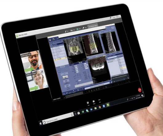
This strategic collaboration’s goal is to enable GE HealthCare to provide a multi-vendor, multi-modality remote scanning solution to healthcare systems and patients around the globe. Digital Expert Access is a real-time, virtual solution that enables collaboration among radiology teams, within a single hospital or across multiple locations.
Introduction: The realm of diagnostic imaging has taken a revolutionary leap with the introduction of Cone Beam ComputedTomography (CBCT). Balancing Safety and Precision: CBCT technology is designed with a commitment to minimizing radiation exposure while maintaining diagnostic accuracy, prioritizing patientsafety.
Introduction: Cone Beam ComputedTomography (CBCT) interpretation is a marriage of artistry and technology, providing a three-dimensional view of the oral and maxillofacial region. Conclusion: Cone Beam ComputedTomography (CBCT) interpretation is a blend of artistry and technology, akin to deciphering a three-dimensional masterpiece.
Introduction: Cone Beam ComputedTomography (CBCT) has emerged as a transformative technology that goes “beyond shadows” in the fields of orthodontics and oral surgery. It offers detailed, three-dimensional insights that transcend traditional imaging methods, revolutionizing treatment planning and patient care.
Introduction: The quest for precision in dental and maxillofacial imaging has found its answer in Cone Beam ComputedTomography (CBCT). Safety and Minimal Radiation Exposure: CBCT technology is designed with patientsafety in mind.
Teleradiology in a flat world Introduction: Cone Beam ComputedTomography (CBCT) technology has transcended its role in dentistry, extending its reach into the broader realm of medical diagnostics. Its digital dimension, with three-dimensional insights, has improved diagnostic precision, treatment planning, and patient care.
Introduction: The advent of Cone Beam ComputedTomography (CBCT) has ushered in a new era in advanced imaging techniques in dentistry. Striking a balance between diagnostic precision and patientsafety is an ongoing concern. This transformative technology has brought both challenges and triumphs to the field.
This blog post explores innovative strategies and technologies aimed at reducing radiation exposure in medical imaging, ensuring patientsafety without compromising diagnostic accuracy. Emphasize the need for a nuanced approach to ensure patientsafety.
Cone Beam ComputedTomography (CBCT) goes beyond traditional panoramas, offering comprehensive insights that are reshaping the way dental professionals diagnose and treat oral conditions. In this article, we’ll explore how CBCT is expanding the horizons of dental practice with its detailed three-dimensional views.
teleradiology India Introduction: In the quiet chambers of medical facilities, a silent revolution is underway, and it’s powered by CT (ComputedTomography) scans. This blog explores how CT scans are silently but profoundly changing the landscape of healthcare, revolutionizing diagnostic precision and patient care.
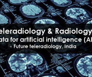
Teleradiology & Radiology data for artificial intelligence (AI) Introduction: The role of a Cone Beam ComputedTomography (CBCT) technologist is vital in the realm of diagnostic imaging. Safety and Radiation Control: Ensuring patientsafety is paramount.
Introduction: The world of digital dentistry has been dramatically transformed by the advent of Cone Beam ComputedTomography (CBCT). Minimal Radiation Exposure: The dental community is dedicated to minimizing radiation exposure, designing CBCT technology to prioritize patientsafety while maintaining diagnostic accuracy.
MRI-Scan-Teleradiology Introduction: Cone Beam ComputedTomography (CBCT) pioneers are at the forefront of innovations that are reshaping the landscape of three-dimensional imaging.
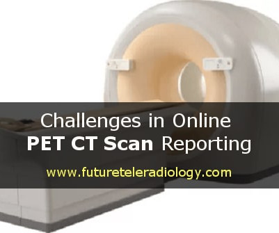
PET-CT-Scan-Reporting-Service Introduction: The world of Cone Beam ComputedTomography (CBCT) technology has evolved from the realm of pixels to the dynamic exploration of volumes. This advancement has revolutionized the field of diagnostic imaging, providing insights that were once unimaginable.
Teleradiology-India Introduction: CT (ComputedTomography) scans offer a window into the human body, but how are these detailed cross-sectional images reconstructed? This blog provides insights into the intricate art and science of CT image reconstruction, shedding light on the technology and expertise that make it all possible.
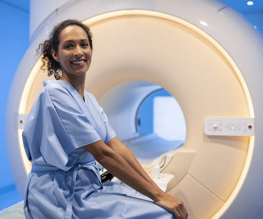
Our centers will pre-screen each patient in order to qualify them as being eligible to safely undergo an MRI. If it is determined that an MRI is not safe for a patient, we often recommend that a ComputedTomography (CT) scan as the best diagnostic alternative. Your safety is our priority.
This is done with the patients on their regular medications so the artifacts due to movements are reduced and clear images are obtained. On the day of surgery, the frame is fixed under local anaesthesia in the radiology suite and a CT scan with the frame is obtained.
We organize all of the trending information in your field so you don't have to. Join 5,000 users and stay up to date on the latest articles your peers are reading.
You know about us, now we want to get to know you!
Let's personalize your content
Let's get even more personalized
We recognize your account from another site in our network, please click 'Send Email' below to continue with verifying your account and setting a password.


Let's personalize your content