Remove Computed Tomography Remove Radiograph Remove Radiography
article thumbnail

X-raying Wisdom: The Precision and Care in Dental Radiography Practices (Gujarat, Jamnagar, Morbi, Mahisagar)

Future Teleradiology

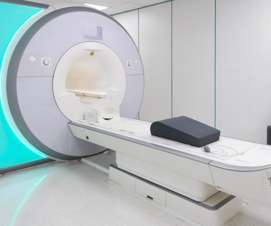
MRI-Scan-Teleradiology Introduction: Dental radiography is an essential component of modern dentistry, offering valuable insights into oral health and guiding treatments with precision. Patient-Centered Approach: Dental radiography starts with the patient. Cone-Beam Computed Tomography (CBCT): CBCT is a game-changer in dental imaging.

article thumbnail

WBCT Indications Series: Midfoot Arthritis

CurveBeam AI

In addition, WBCT better quantifies 3 the structural deformity of Chopart, talonavicular, and calcaneocuboid joints when compared to conventional radiography and non-weight bearing computed tomography images. However, the severity of deformity and involved joints were difficult to determine on plain radiographs alone.

Insiders

Sign Up for our Newsletter

This site is protected by reCAPTCHA and the Google Privacy Policy and Terms of Service apply.

Trending Sources

article thumbnail

New Campaign From the American Society of Radiologic Technologists Advocates for Profession to "Be Seen"

Imaging Technology

To highlight the profession, ASRT has partnered with medical imaging professional and actor, Michael Benzaia.

article thumbnail

WBCT Indications Series: Lisfranc

CurveBeam AI

Diagnosis The diagnosis of Lisfranc injuries may be challenging on plain radiographs alone. Radiographs were indeterminate. Comparative assessment of midfoot osteoarthritis diagnostic sensitivity using weightbearing computed tomography vs weightbearing plain radiography. She underwent ORIF of her Lisfranc injury.

article thumbnail

WBCT Indications Series: PCFD

CurveBeam AI

A weight bearing CT scan can: Provide an assessment of important anatomical markers of pronounced hindfoot deformity and peritalar subluxation (PTS), difficult to visualize on conventional two-dimensional radiographs 1. Weightbearing Computed Tomography for Assessment of Foot and Ankle Deformities: The Iowa Experience.

article thumbnail

Inside the Jaw: A Day in the Life of a Dental X-ray Technologist (Gujarat, Anand, Chhota Udaipur, Banaskantha)

Future Teleradiology

Introduction: Dental X-ray technologists, also known as dental radiographers, are the unsung heroes behind the scenes of every successful dental diagnosis and treatment. Patient Interaction: Patient Comfort: Dental radiographers are responsible for ensuring the patient’s comfort and alleviating any anxiety.

X-ray 52
article thumbnail

Lisfranc Fracture Dislocation

Global Radiology CME

A) AP radiograph of Lisfranc Fracture Dislocation demonstrates the circled “fleck sign” or Lisfranc ligament avulsion fracture fragment. (B) C) The lateral radiograph notes with a circle, the dorsal sub dislocation of the metatarsal base. Radiographs should be repeated after two weeks to ensure surgery is unnecessary.