This site uses cookies to improve your experience. To help us insure we adhere to various privacy regulations, please select your country/region of residence. If you do not select a country, we will assume you are from the United States. Select your Cookie Settings or view our Privacy Policy and Terms of Use.
Cookie Settings
Cookies and similar technologies are used on this website for proper function of the website, for tracking performance analytics and for marketing purposes. We and some of our third-party providers may use cookie data for various purposes. Please review the cookie settings below and choose your preference.
Used for the proper function of the website
Used for monitoring website traffic and interactions
Cookie Settings
Cookies and similar technologies are used on this website for proper function of the website, for tracking performance analytics and for marketing purposes. We and some of our third-party providers may use cookie data for various purposes. Please review the cookie settings below and choose your preference.
Strictly Necessary: Used for the proper function of the website
Performance/Analytics: Used for monitoring website traffic and interactions
Diagnostic radiology MRI Safety A new CPT subsection has been established for reporting six new codes describing MR safety services, including implant or foreign body evaluation, safety consultation, electronics preparation, and implant positioning or immobilization. PC-1.97 $120.01 $63.72 64467 unilateral; by infusion(s) G-6.86
Women in racial and ethnic minority backgrounds are less likely to be provided same-day diagnostic breast imaging services, despite such services being available, according to research published February 18 in Radiology. Additional imaging and possibly image-guided biopsy are recommended for women who have an abnormal screening mammogram.
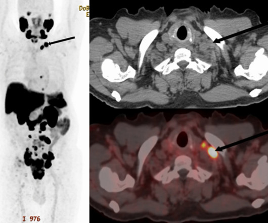
PET/CT scans with an experimental prostate-specific membrane antigen (PSMA) imaging agent can identify supraclavicular nodal metastasis in newly diagnosed prostate cancer patients, researchers have reported. For the analysis, the researchers included 240 patients who underwent scans for primary staging of newly diagnosed disease.
Chiao, a former NASA astronaut and ISS commander, was part of the Advanced Diagnostic Ultrasound in Microgravity(ADUM) project. Chiao told personal stories of his time in space, along with how ultrasound findings have helped in developing health measures and diagnostic protocols for spaceflight.
Technetium-99m (Tc-99m) methyl diphosphonate (MDP) bone scans are a potentially viable noninvasive option for diagnosing calciphylaxis, according to a team at the University of Massachusetts in Worcester, MA. The team assessed the potential diagnostic utility of bone scans in calciphylaxis based on a review of the literature.
Point-of-care ultrasound (POCUS) can be a useful tool for diagnosing "retained products of conception" in women, suggest findings published January 13 in the American Journal of Emergency Medicine. Once diagnosed, women are treated via dilation and curettage, the drug misoprostol, or ongoing follow-up. and specificity would be 90.4%.
Lymphoscintigraphy (LSG) is recommended but seldom used to diagnose lymphedema in real-world settings in the U.S., The finding comes despite guidelines recommending LSG as the diagnostic test of choice and underlines the need for a better diagnostic test, wrote lead author Tina Moon, MD, of Tufts Medical Center in Boston and colleagues.
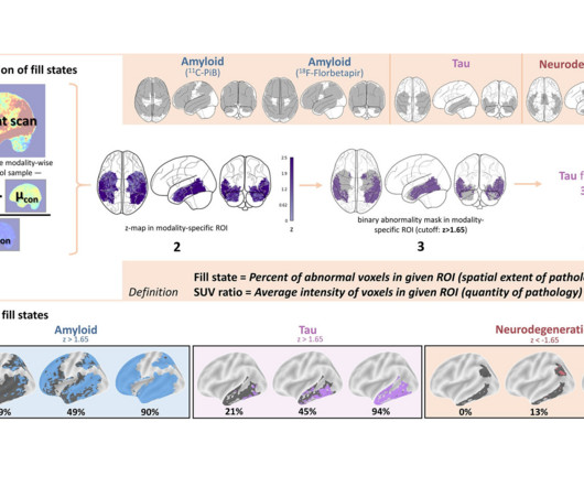
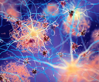
Researchers in Germany have proposed a new approach derived from brain PET imaging for diagnosing and staging Alzheimers disease, according to a study published March 25 in Radiology.
FDG-PET imaging shows promise for use as a diagnostic criterion for neurosarcoidosis, with a recent case series illustrating the approach was effective when gold-standard approaches were not, according to a group of researchers in Berlin. “To
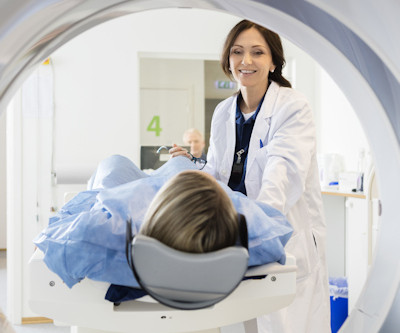
Diagnostic Imaging is a great tool for your medical professional to use to detect issues sooner rather than later. There are several types of diagnostic imaging available today; each one used to visualize the internal structures of the body to assist doctors in diagnosis and treating various diseases and medical conditions.
“Ultrasound contrast agent use can be ubiquitous in interventional practice and can solve many logistical or clinical diagnostic problems,” he said. In ultrasound, contrast use has been shown to improve differentiation of liver lesions and the diagnostic performance of O-RADS in assessing ovarian lesions among other examples.
PET/MRI imaging shows promise in diagnosing fevers or inflammation of unknown origin and may have advantages over PET/CT, according to a study published January 3 in the European Journal of Radiology. Fever of unknown origin (FUO) is defined as a temperature higher than 38.3 °C
But evidence as to how this type of upgrade impacts various pathways for diagnosing prostate cancer is unclear. To address the knowledge gap, the team evaluated the benefit of using PI-RADS upgrading rules with MRI-directed diagnostic sequences.
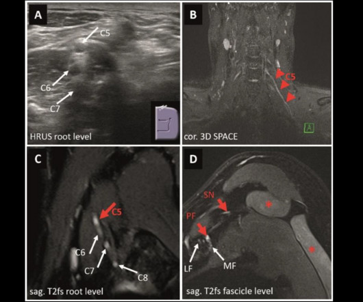
MRI and ultrasound have tradeoffs in diagnosing peripheral neuropathies of the upper extremity, a study published March 4 in Radiology found. High-resolution imaging provides important pathomorphological information in diagnosing peripheral nerve disorders. MRN and HRUS are two imaging techniques that have shown promise in this area.
“Our study showed that the availability of PET/CT prior to the [percutaneous needle lung biopsy] improves the diagnostic biopsy rates,” wrote lead author Konstantinos Stefanidis, MD, of King’s College Hospital in London, and colleagues. Patient with extensive lung and pleural disease. (2a) Image courtesy of the European Journal of Radiology.
A thyroid ultrasound imaging database could spur the development of more effective diagnostic and treatment models for related diseases, according to research published January 23 in Ultrasound in Medicine & Biology. Ultrasound is a first-line method in the screening and diagnosing of thyroid nodules.
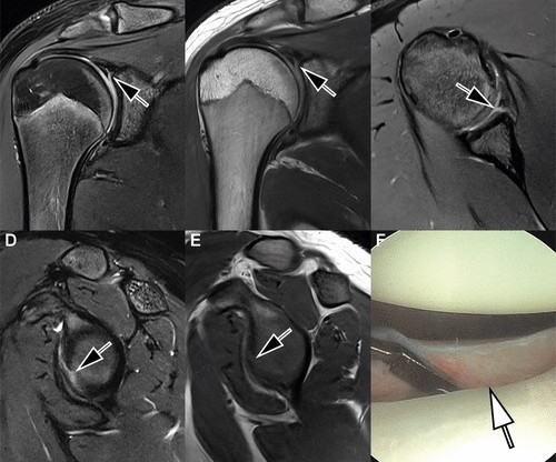
DL algorithms have been developed over the past five years to reduce these motion artifacts, yet evidence on their diagnostic performance is scarce, the group noted. All seven readers correctly diagnosed the arthroscopy-validated findings. (F) F) An arthroscopic photograph shows the anteroinferior labral tear (arrow).
Her team studied how an AI-guided POCUS system can help diagnose tuberculosis in patients with respiratory symptoms. Suttels highlighted lung POCUS being connected to a smartphone to make diagnoses and being sputum-free. Image courtesy of Vronique Suttels, PhD.
Virtual/augmented reality (VR/AR) headsets show promise for helping clinicians interpret CT exams -- even for conditions such as diverticulitis, which can be tricky to diagnose, researchers have reported. The use of these headsets instead of desktop displays has been explored for both image-guided procedures and diagnostic interpretation.
A team led by Seyed-Ali Sadegh-Zadeh, PhD, from Staffordshire University in England found that its machine-learning model achieved high performance in diagnosing heart tumors, including a near-perfect area under the curve (AUC) score. The researchers highlighted that machine learning techniques could lead to improved diagnostic performance.
12, 2025 Konica Minolta Healthcare Americas, has published a case study by clinicians in the pulmonary and radiology departments at ASST Fatebenefratelli Sacco (Milan, Italy) demonstrating the use of Dynamic Digital Radiography (DDR) to help definitively diagnose diaphragm dysfunction. tim.hodson Fri, 02/14/2025 - 15:14 Feb.12,
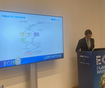
In his talk, Niccol Finardi from the University of Milan in Italy presented his teams research, demonstrating CEUS high diagnostic accuracy at different time points after spleen trauma occurs, ranging from within 48 hours to 60 days follow-up. One patient dropped out of the study due to the critical nature of their injury.
ACR is teaming with Belgium-based Icometrix, which is providing its brain MRI software to facilities participating in the Alzheimers Network for Treatment and Diagnostics.
ChatGPT-4 has greater diagnostic accuracy than ChatGPT-3.5 However, the researchers pointed out that it remains to be seen how upgrades in ChatGPT improve performance in providing diagnoses in radiology cases. The team investigated the respective diagnostic accuracy of ChatGPT-3.5 Finally, diagnostic accuracy increased by 17.3%
When it comes to your health, selecting the right diagnostic imaging center is an important decision. State-of-the-Art Technology Not all imaging centers offer the same equipment, and newer technology often leads to more precise imaging and quicker diagnoses. Here are some key factors to keep in mind when making your choice.
Background: The increased utility and accessibility of point-of-care ultrasound (POCUS) has allowed clinicians the freedom to rethink their diagnostic approach for many common diseases, including peritonsillar abscess (PTA). Question: How well does the US perform in diagnosing PTA compared to CT, needle aspiration, or Incision and Drainage?
SimonMed Imaging has inked a research collaboration with Oxford Brain Diagnostics to explore using cortical disarray measurement (CDM) to better diagnose neurodegenerative diseases. The companies aim to demonstrate the accuracy and efficacy of CDM compared with other diagnostic methods including standard MRI and PET.
Chest x-ray is used to diagnose RDS, but LUS has emerged as a reliable bedside tool for monitoring respiratory conditions in preterm infants. Specificity 50% 91% Diagnostic accuracy 80.1%
Axillary scanning in women undergoing a diagnostic breast ultrasound has minima. New breast elastography algorithm avoids false-negative cases Ultrasound, AI method diagnoses breast lumps without experts Initial results for SOLUS trial presented at ECR
diagnostic support technology uses AI to analyze CT scans, x-rays, and pathology slides, supporting clinicians in diagnosing medical conditions earlier and with better accuracy, improving clinical decision-making and patient outcomes. operations and growing its customer base. Harrison.ai
They also spearheaded the development of guidelines for the American College of Radiology (ACR), recognizing the role that imaging had in diagnosing COVID-19 in patients. The guidelines included best practices for imaging with CT versus chest x-ray in diagnosing COVID, as well as whether imaging is necessary at all in some cases.
A team led by Mark Slidell, MD, from Johns Hopkins University in Baltimore, MD, found that CT remains the most used imaging method for diagnostic pediatric appendicitis in hospitals that don’t participate in the National Surgical Quality Improvement Program - Pediatrics (NSQIP-P). NSQIP-P hospital 71.7% Non-NSQIP-P hospital 18.8%
Boussoussou and colleagues conducted a study that assessed the diagnostic efficacy of PCCT for this indication, using ICA as a reference. Photon counting coronary CT (PCCT) angiography represents a groundbreaking approach to evaluating coronary stenosis," the group concluded.
Centers for Medicare and Medicaid Services (CMS) proposed a new reimbursement plan July 10 for diagnostic PET scans that would provide separate payments for radiopharmaceuticals, as well as an extra payment for hospitals when they use domestically produced technetium-99m (Tc-99m).
Using direct radiologic image inputs can improve the diagnostic accuracy of large language models, according to research published July 9 in Radiology. Suh and co-authors studied the performance of these two models in generating differential diagnoses at different temperatures.
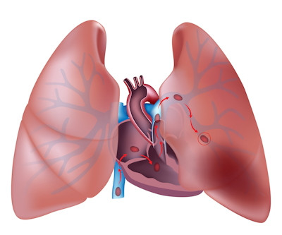
Remy-Jardin and colleagues conducted a study that compared performance of traditional CT to PCCT for diagnosing acute PE. Performance comparison of traditional CT to PCCT for diagnosing acute pulmonary embolism Measure Group 1 (dual-energy protocol) Group 2 (multienergy protocol) Percent reduction Acquisition time 4 seconds 0.9
Chinese clinicians have provided evidence in a “real-world study” that shows amyloid PET imaging is effective for diagnosing and managing patients with Alzheimer’s disease, according to a study published February 8 in Alzheimer’s and Dementia. of patients, noted lead author Ke-Liang Chen, MD, a neurologist at Fudan University.
Under the new policy, CMS will unpackage and pay separately for diagnostic radiopharmaceuticals with per-day costs exceeding $630, removing financial barriers that have long hindered patient access to essential nuclear medicine diagnostic procedures.
SINGAPORE – MR elastography is a leading noninvasive approach for imaging liver fibrosis, yet a new technique known as “3D MRE” promises to open new opportunities for diagnosing chronic liver disease, according to a leading expert in the field.
Conebeam breast CT (CBBCT) has superior diagnostic performance than that of mammography in small studies, according to research published January 8 in the European Journal of Radiology. CBBCT in recent years has garnered interest among radiologists as another way to diagnose breast cancer.
We organize all of the trending information in your field so you don't have to. Join 5,000 users and stay up to date on the latest articles your peers are reading.
You know about us, now we want to get to know you!
Let's personalize your content
Let's get even more personalized
We recognize your account from another site in our network, please click 'Send Email' below to continue with verifying your account and setting a password.


Let's personalize your content