Remove Diagnose Remove Diagnostic Remove Global Radiology Remove Radiographer
article thumbnail

Lisfranc Fracture Dislocation

Global Radiology CME

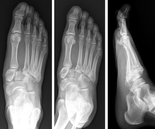
A) AP radiograph of Lisfranc Fracture Dislocation demonstrates the circled “fleck sign” or Lisfranc ligament avulsion fracture fragment. (B) C) The lateral radiograph notes with a circle, the dorsal sub dislocation of the metatarsal base. of all diagnosed fractures. Trauma due to falling off a roof. Xray of the Week Figure 1.

article thumbnail

Lens Subluxation

Global Radiology CME

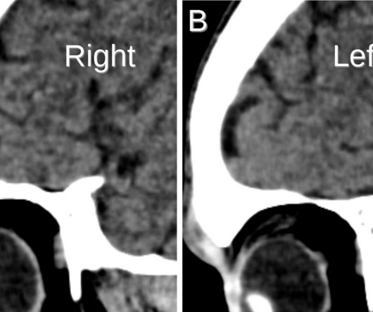
Lens subluxation can be diagnosed by ultrasound which shows deviation of the lens (Fig. Radiographics. Trauma-Induced Bilateral Ectopia Lentis Diagnosed with Point-of-Care Ultrasound. Dr. Rice's passion for state of the art radiology and teaching includes acting as a guest lecturer at UCLA. 1, 2) [4]. 2006;10(4):345-350.

Insiders

Sign Up for our Newsletter

This site is protected by reCAPTCHA and the Google Privacy Policy and Terms of Service apply.

Trending Sources

article thumbnail

Lens Dislocation

Global Radiology CME

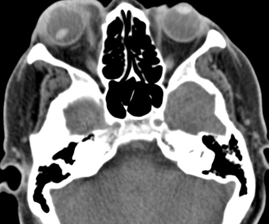
CT is the most commonly used diagnostic modality in lens dislocation and can clearly show lens displacement [4]. Ultrasound has been shown to rapidly diagnose lens dislocation as it can visualize internal structures of the globe [2, 7]. Radiographics. He plans on pursuing a residency in Diagnostic Radiology.

article thumbnail

Porencephaly

Global Radiology CME

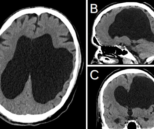
Porencephaly is readily diagnosed via imaging. Diagnostic Imaging: Obstetrics, Elsevier, 2016, pp. Jay Vora is a medical student at Edward Via College of Osteopathic Medicine (VCOM) – Virginia and plans to pursue a residency in diagnostic radiology. Textbook of Fetal Abnormalities, Elsevier, 2007, pp.