This site uses cookies to improve your experience. To help us insure we adhere to various privacy regulations, please select your country/region of residence. If you do not select a country, we will assume you are from the United States. Select your Cookie Settings or view our Privacy Policy and Terms of Use.
Cookie Settings
Cookies and similar technologies are used on this website for proper function of the website, for tracking performance analytics and for marketing purposes. We and some of our third-party providers may use cookie data for various purposes. Please review the cookie settings below and choose your preference.
Used for the proper function of the website
Used for monitoring website traffic and interactions
Cookie Settings
Cookies and similar technologies are used on this website for proper function of the website, for tracking performance analytics and for marketing purposes. We and some of our third-party providers may use cookie data for various purposes. Please review the cookie settings below and choose your preference.
Strictly Necessary: Used for the proper function of the website
Performance/Analytics: Used for monitoring website traffic and interactions
Women in racial and ethnic minority backgrounds are less likely to be provided same-day diagnostic breast imaging services, despite such services being available, according to research published February 18 in Radiology. Additional imaging and possibly image-guided biopsy are recommended for women who have an abnormal screening mammogram.
The COVID-19 pandemic prompted an increase in the rates of negative mammograms in both screening and diagnostic settings, a study published November 14 in the Journal of Radiology Nursing found. They also found a decrease in the proportion of negative diagnosticmammograms.
They also spearheaded the development of guidelines for the American College of Radiology (ACR), recognizing the role that imaging had in diagnosing COVID-19 in patients. The guidelines included best practices for imaging with CT versus chest x-ray in diagnosing COVID, as well as whether imaging is necessary at all in some cases.
Komen urged quick passage of legislation introduced in Pennsylvania to eliminate out-of-pocket costs for women for necessary diagnostic breast cancer imaging. Gina Curry (D-Delaware) and would eliminate costs for women for supplemental imaging such as breast MRIs and ultrasounds.
ChatGPT-4 outperformed human clinicians in determining pretest and post-test disease probability after a negative test result involving chest radiographs and mammograms, according to a research letter published December 11 in JAMA Network Open.
christine.book Tue, 05/21/2024 - 10:36 May 21, 2024 — According to a newly-published study of nearly 5,000 screening mammograms interpreted by an FDA-approved AI algorithm, patient characteristics such as race and age influenced false positive results. Nguyen, M.D.
It is estimated that 1 out of every 8 women in the United States will be diagnosed with breast cancer in her lifetime. One question our technologists are asked frequently is, “What’s the difference between a diagnosticmammogram and a screening mammogram?” first appeared on Clermont Radiology.
Komen has urged quick passage in Arizona of legislation that would eliminate patient out-of-pocket costs for diagnostic and supplemental breast imaging. House Bill 2411 was introduced in the state by Representative David Cook (R-Globe) and includes eliminating costs for patients for MRI, ultrasound, and diagnosticmammograms.
“We're excited to introduce advanced workstation features for our flagship solution, ProFound Detection, aimed at further improving and facilitating radiologists' interpretation of mammograms within their workstation,” said Dana Brown , President and CEO of iCAD.
CNNs have been used in AI research to classify breast cancer disease status and predict therapeutic responses based on diagnostic images of the primary breast tumor. The researchers highlighted that CNNs require little preprocessing compared with other image classification algorithms. DEED Attribution-NonCommercial-NoDerivs 4.0 International.
The American Cancer Society recommends starting annual mammogram screenings at age 40. The list below are imaging exams used to diagnose breast cancer. Mammogram Screening Mammogram: Screening mammograms take 2 or more images of each breast. The list below are imaging exams used to diagnose breast cancer.
In addition, the ACR recommends that women diagnosed with breast cancer prior to age 50 or with a personal history of breast cancer and dense breasts should have annual supplemental breast MRI. Food and Drug Administration (FDA) requirement that all women having mammograms receive notice that their breasts are dense or not dense.
The earlier breast cancer is diagnosed, the easier it is to treat. The earlier breast cancer is detected through diagnostic imaging, the better chance there is for successful treatment with surgery, radiation therapy, or chemotherapy. To that end, you should be aware of the common signs and symptoms of breast cancer. this year alone.
Artificial Intelligence (AI): Revolutionizing Radiology in 2025 AI continues to make waves in radiology, offering improved diagnostic accuracy and efficiency. These facilities are adopting cutting-edge imaging technology, enabling faster and more accurate diagnoses. Read more about AI advancements in radiology here.
A team of AI and medical specialists working with or for Google Research and Google DeepMind, has developed an AI based system designed to judge the confidence level of existing AI systems used for analyzing medical scans as a means of improving analysis of diagnostic tools, such as mammograms or chest X-rays.
A combination of canceled elective screenings and procedures, staff and PPE shortages, office closures, and personal health concerns all contributed to a decline in the number of routine mammograms provided for at-risk women. The result is a rise in breast cancer diagnoses. Why do I need a Mammogram Every Year?
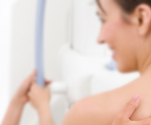
Screening Mammograms have proven to be essential for the early detection of breast cancer. What Is A Screening Mammogram? A mammogram is an examination that uses a special low dose X-Ray machine to evaluate breast tissue. The mammogram unit is designed specifically for breast imaging. Are Screening Mammograms Risky?
Teleradiology: Bridging the Gap in Underserved Areas Teleradiology plays a pivotal role in expediting cancer diagnoses, particularly in rural or underserved regions. These benefits collectively enhance the overall performance of healthcare facilities by streamlining operations and ensuring high-quality diagnostic services.
As a teleradiology company, we specialize in bridging this gap by offering high-quality diagnostic imaging interpretation, ensuring rural healthcare providers can deliver top-tier care to their patients. Radiologists must inform patients if they have dense breast tissue, a factor that can obscure mammogram results and increase cancer risks.
That is, it is diagnosed after a test whose result was negative and before the next evaluation. Conversely, if the number of tumors diagnosed after screening is low, it indicates that the program is fulfilling its function: to detect this disease at an early stage. per thousand people, while in the second it was 0.93.
Teleradiology-in-Flat-World Introduction : In the realm of modern medicine, diagnostic tools such as X-Rays, mammograms, and CAT scans play a crucial role in identifying and understanding various health conditions. Medical malpractice claims can arise from misdiagnoses and improper use of diagnostic tools.
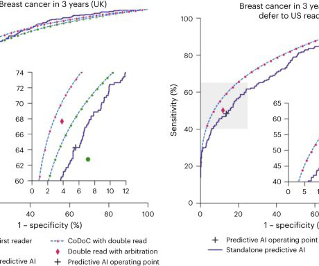
Using mammography-based deep learning models may improve the accuracy of breast cancer risk assessment and can also lead to earlier diagnoses. “About 1 in 10 women develop breast cancer throughout their lifetime,” said study author Andreas D. Lauritzen , Ph.D., A variety of AI tools exist to aid in detecting cancer risk.
Breast cancer is the most commonly diagnosed form of cancer in American women. National initiatives are in place to urge women everywhere to get regular screenings, but there are some real disparities that exist when it comes to who is getting mammograms and how often.
mtaschetta-millane Mon, 07/29/2024 - 11:16 July 29, 2024 — Lunit, a leading provider of AI-powered solutions for cancer diagnostics and therapeutics, announced the implementation of its AI-powered breast cancer detection solution, Lunit INSIGHT MMG , in Qatar's national breast cancer screening program.
Low-dose positron emission mammography (PEM) is a novel molecular imaging technique that provides improved diagnostic performance at a radiation dose comparable to that of mammography. Low-dose PEM offers potential clinical uses in both screening and diagnostic settings, according to Dr. Freitas.
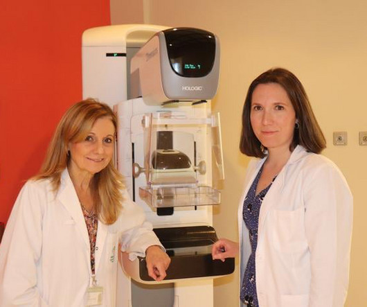
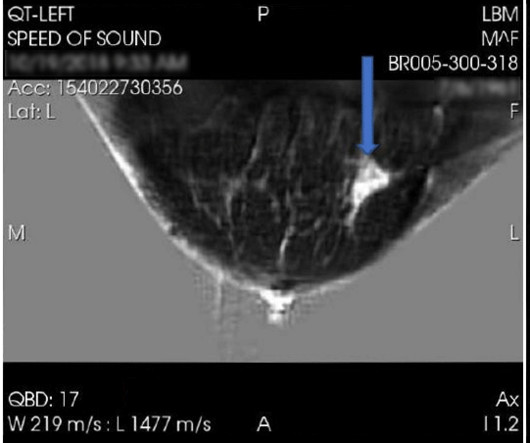
a medical device company engaged in research, development, and commercialization of innovative body imaging systems, announced positive data regarding the diagnostic performance of QTI’s Breast Acoustic CT Scans for mass detection from its second blinded multi-reader multi-case study.
milla1cf Thu, 12/14/2023 - 08:21 December 14, 2023 — Lunit , a leading provider of AI-powered solutions for cancer diagnostics and therapeutics, today announced its proposal to acquire Volpara Health Technologies Ltd. , Lunit's acquisition of Volpara signifies a pivotal moment in our commitment to advancing global cancer diagnostics.
In addition to mammograms and ultrasounds, women should know about the various other breast screenings that are performed. Diagnostic Mammography Diagnosticmammograms may be performed when a woman (or man) has presented with some type of symptom that requires further examination.
Beyond diagnostics, AI has played a pivotal role in drug discovery, streamlining clinical trials, and personalizing patient interventions. In the United States alone, it is estimated that around 40 million mammograms were performed each year. Mammograms face challenges due to factors like breast density, leading to missed cancer cases.
28, 2024 — Rezolut, LLC recently debuted its latest offering for patients during their annual mammogram, SecondReadAI powered by Lunit. Leveraging deep learning techniques, the system analyzes mammography images, providing radiologists with valuable insights and supporting them in their diagnostic process.
The latest in a series of such research competitions that RSNA has conducted since 2015, this challenge tasked participants with developing artificial intelligence (AI) models that can accurately detect breast cancer from mammography images, potentially assisting radiologists in making more accurate and timely diagnoses.
The study’s limitations include the potential misclassification of diagnosticmammograms as screening and the inability to adjust for certain breast cancer risk factors. The post Breast Cancer Awareness Month Kicks Off Now: The Latest in Breast Cancer Studies first appeared on Vesta Teleradiology.
You’re more likely to be diagnosed with breast cancer than any other cancer (besides skin cancer). That decline has been attributed, in large part, to annual screening mammograms. Each year, during your mammogram appointment, a full assessment is done to determine if you have one or multiple risk factors.
All of the annual scheduled services such as mammograms can now be scheduled, as well as imaging prescribed by physicians for the care of their patients. Patients may also schedule mammograms directly at our facilities in these same locations. What this means for our community and region is important.
When mammography is carried out on an individual without any symptoms, it is called screening mammography whereas if carried out on an individual with symptoms, it is called diagnostic mammography in which additional views have to be carried out. If the breast is dense on the mammogram, an ultrasound must also be carried out.
With an X-ray, Ultrasound, Mammogram and CT scan at our disposal, we needed to have a radiologist who could read all these modalities, and give us results in the shortest time possible to enable us to give the best medical care possible” she says. She has since received definitive treatment and is doing well.
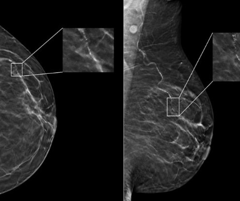
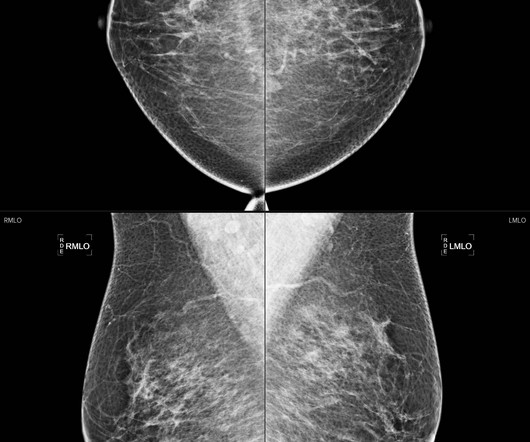
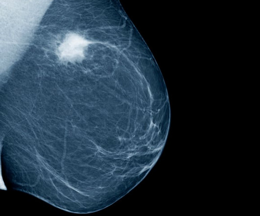
Women with dense breasts are BOTH more likely to develop breast cancer and more likely to have that cancer missed on a mammogram [5] Fig. 1 – Cancer on a mammogram of a fatty vs a dense breast What is Dense Breast Tissue? Breast density is determined through a mammogram and described as one of four categories (Fig.
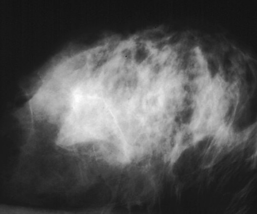
A) Mammogram MLO view. Mammogram CC view. A mammogram demonstrated focal asymmetries involving most of the anterior and mid right breast with diffuse skin thickening, trabecular coarsening, increased overall density, and enlarged right axillary lymph nodes. What is the diagnosis? Xray of the Week Figure 1. Breast Cancer.
Doctors use imaging tests to see inside a patient’s body and diagnose their illnesses and injuries. A doctor will often use a diagnostic imaging test to check patients for signs of cancer. Diagnosis The diagnosis of cancer is often first detected through a diagnostic screening. This is known as cancer staging.
Teleradiology for Indian Health Services As a premier teleradiology company, Vesta understands the critical importance of reliable and efficient diagnostic imaging services, especially in regions facing shortages. With our state-of-the-art technology and a team of highly skilled U.S.
When it comes to accurate diagnoses and effective patient care, getting a second opinion on imaging results can make all the difference. Our abdominal imaging specialists ensure accurate diagnoses for conditions like pancreatic cancer or complex GI issues. Why Choose a Teleradiology Partner for Second Opinions?
An MCQ that asks the learner to recognize benign dermal calcifications on a mammogram does not test the learner’s problem-solving ability or ability to communicate the findings to a patient. That is, they should fall into the same category as the correct option (all diagnoses, tests, treatments, prognoses, disposition alternatives).
IR came to life through the combination of the creative thinking and technical skills of diagnostic radiologists and angiogiographers. (11) It was only taken seriously after a large ovarian cyst in a female patient was diagnosed, as at first Donald’s idea was ridiculed. (14) Their work gave rise to the modern MRI scanners we use today.
milla1cf Fri, 04/12/2024 - 21:00 April 12, 2024 — GE HealthCare , a leader in breast health technology and diagnostics, will feature its latest breast cancer detection technology during the 2024 Society of Breast Imaging Symposium in Montreal, Canada, April 11-14, 2024.
We organize all of the trending information in your field so you don't have to. Join 5,000 users and stay up to date on the latest articles your peers are reading.
You know about us, now we want to get to know you!
Let's personalize your content
Let's get even more personalized
We recognize your account from another site in our network, please click 'Send Email' below to continue with verifying your account and setting a password.


Let's personalize your content