This site uses cookies to improve your experience. To help us insure we adhere to various privacy regulations, please select your country/region of residence. If you do not select a country, we will assume you are from the United States. Select your Cookie Settings or view our Privacy Policy and Terms of Use.
Cookie Settings
Cookies and similar technologies are used on this website for proper function of the website, for tracking performance analytics and for marketing purposes. We and some of our third-party providers may use cookie data for various purposes. Please review the cookie settings below and choose your preference.
Used for the proper function of the website
Used for monitoring website traffic and interactions
Cookie Settings
Cookies and similar technologies are used on this website for proper function of the website, for tracking performance analytics and for marketing purposes. We and some of our third-party providers may use cookie data for various purposes. Please review the cookie settings below and choose your preference.
Strictly Necessary: Used for the proper function of the website
Performance/Analytics: Used for monitoring website traffic and interactions
Diagnostic radiology MRI Safety A new CPT subsection has been established for reporting six new codes describing MR safety services, including implant or foreign body evaluation, safety consultation, electronics preparation, and implant positioning or immobilization. PC-6.47 $525.63 $209.28 PC-14.56 $8,508.75 $470.97 PC-14.56 $8,508.75 $470.97
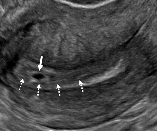
Point-of-care ultrasound (POCUS) can be a useful tool for diagnosing "retained products of conception" in women, suggest findings published January 13 in the American Journal of Emergency Medicine. Once diagnosed, women are treated via dilation and curettage, the drug misoprostol, or ongoing follow-up.
A thyroid ultrasound imaging database could spur the development of more effective diagnostic and treatment models for related diseases, according to research published January 23 in Ultrasound in Medicine & Biology. Ultrasound is a first-line method in the screening and diagnosing of thyroid nodules.
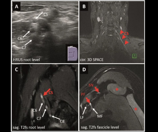
MRI and ultrasound have tradeoffs in diagnosing peripheral neuropathies of the upper extremity, a study published March 4 in Radiology found. High-resolution imaging provides important pathomorphological information in diagnosing peripheral nerve disorders. Images and caption courtesy of the RSNA. Sensitivity 91.6%
While the use of ultrasound for pediatric appendicitis imaging has increased, the modality -- as well as MRI -- remains underutilized in this area, according to research published August 22 in the Journal of Pediatric Surgery. Ultrasound alone was used in 53.3% Ultrasound alone was used in 53.3% NSQIP-P hospital 71.7%
Contrast-enhanced ultrasound (CEUS) could be a suitable alternative to CT for early detection of complications in splenic trauma thats nonoperatively managed, according to a study presented on February 28 at ECR 2025. Finardi, however, highlighted that timing for such management has not been established.
Dynamic contrast-enhanced ultrasound (CEUS) is a useful tool for interventions, according to a March 3 presentation given at ECR 2024. Ultrasound contrast agent use can be ubiquitous in interventional practice and can solve many logistical or clinical diagnostic problems,” he said.
Lymphoscintigraphy (LSG) is recommended but seldom used to diagnose lymphedema in real-world settings in the U.S., The finding comes despite guidelines recommending LSG as the diagnostic test of choice and underlines the need for a better diagnostic test, wrote lead author Tina Moon, MD, of Tufts Medical Center in Boston and colleagues.
A computer-aided diagnostic (CAD) system improves the diagnostic performance of breast ultrasound, according to findings published October 6 in Heliyon. Ultrasound is the go-to modality for diagnostic breast imaging after suspicious mammography findings. However, conventional ultrasound isn’t without its faults.
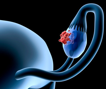
An ultrasound test can identify 96% of ovarian cancers in postmenopausal women, researchers have reported. Early diagnosis of ovarian cancer is vital, and we are pleased to see this research demonstrate that there are more accurate ways of using ultrasound," Sundard and colleagues said in a statement released by the university.
Diagnostic Imaging is a great tool for your medical professional to use to detect issues sooner rather than later. There are several types of diagnostic imaging available today; each one used to visualize the internal structures of the body to assist doctors in diagnosis and treating various diseases and medical conditions.
Studies to be presented in Chicago will explore ultrasound’s clinical applications in musculoskeletal, pediatric, abdominal, and women's imaging among other applications. Furthermore, ultrasound's use as a supplemental tool will be explored, including for breast cancer detection and follow-up imaging. And let’s not forget about AI.
Rural, older, and poorer children face barriers in obtaining an ultrasound exam for acute appendicitis, according to research published October 17 in the Journal of Surgical Research. It also found that they are less likely to undergo ultrasound at hospitals that have ultrasound resources. emergency departments.
Lung ultrasound (LUS) scoring is reliable for determining whether preterm newborns need surfactant for suspected respiratory distress syndrome (RDS), a study published February 19 in Pediatrics & Neonatology found. It also compared ultrasound scores with chest x-ray scores to predict the need for surfactant administration.
“Living” and “viable” should be avoided in first-trimester ultrasound lexicon when imaging pregnant women, according to new guidelines introduced August 27 by the Society of Radiologists in Ultrasound (SRU), published in Radiology.
Axillary scanning in women undergoing a diagnostic breast ultrasound has minima. Read more on AuntMinnie.com Related Reading: Systemic factors linked to incomplete breast imaging follow-up Could ultrasound be used before DBT in focal breast complaints?
Background: The increased utility and accessibility of point-of-care ultrasound (POCUS) has allowed clinicians the freedom to rethink their diagnostic approach for many common diseases, including peritonsillar abscess (PTA). Clinical evaluation may prove difficult since many PTA and peritonsillar cellulitis symptoms overlap.
12, 2025 Konica Minolta Healthcare Americas, has published a case study by clinicians in the pulmonary and radiology departments at ASST Fatebenefratelli Sacco (Milan, Italy) demonstrating the use of Dynamic Digital Radiography (DDR) to help definitively diagnose diaphragm dysfunction. tim.hodson Fri, 02/14/2025 - 15:14 Feb.12,
A deep-learning method using endoscopic ultrasound images could detect pancreatic neuroendocrine neoplasms, according to research published October 17 in Gastrointestinal Endoscopy. While endoscopic ultrasound is sensitive in detecting pancreatic neuroendocrine neoplasms, the modality is operator-dependent and time-consuming.
While services for breast and lung cancer screening were temporarily halted, imagers in x-ray, lung ultrasound, and PET/CT were busy examining patients who presented with COVID-19. The guidelines included best practices for imaging with CT versus chest x-ray in diagnosing COVID, as well as whether imaging is necessary at all in some cases.
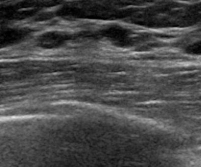
milla1cf Tue, 04/04/2023 - 17:24 April 4, 2023 — Ultrasound is an effective standalone diagnostic method in patients with focal breast complaints, according to a study published in Radiology , a journal of the Radiological Society of North America ( RSNA ). The quality of ultrasound images has also vastly improved in recent years.
At TRA Medical Imaging, we’re here to help you understand how diagnostic imaging can aid in identifying and treating these cold-weather ailments. Who to See: Start with a primary care physician (PCP) or urgent care provider for a thorough evaluation, including a physical exam and initial diagnostic tests.
This was a case of the radiology practice miscoding an ultrasound exam conducted on the pregnant patient at 41 weeks that showed low amniotic fluid -- which resulted in the scan being sent "to nowhere," rather than to the mother's midwife. The baby died. Ferreira had reportedly retired three months prior to the trip.
A team led by Seyed-Ali Sadegh-Zadeh, PhD, from Staffordshire University in England found that its machine-learning model achieved high performance in diagnosing heart tumors, including a near-perfect area under the curve (AUC) score. The researchers highlighted that machine learning techniques could lead to improved diagnostic performance.
A new computer-based diagnostic tool combines ultrasound imagery with specific clinical indicators to evaluate the risk of moderate-to-severe renal fibrosis.
Computer-aided diagnosis (CAD) can enhance the diagnostic performance of breas. Ultrasound, AI method diagnoses breast lumps without experts Ultrasound, AI method diagnoses breast lumps without experts
On Friday, our hospital received 7 Butterfly iQ+ ultrasound devices, which will be indispensable in our work with the pediatric patients," he said in a Facebook post on October 21. Stanislav Rebenkov, MD, has been moved by the support and generosity of the global radiology community. "On
Read more on AuntMinnie.com Related Reading: AI proves comparable to rads for assessing BI-RADS 4 breast lesions ABUS, CEUS could help predict breast cancer treatment response Is axillary scanning needed in diagnostic breast ultrasound?
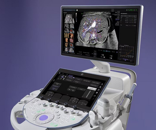
28, 2025 GE HealthCare recently announced it has received 510(k) clearance from the United States Food and Drug Administration (FDA) for the updated portfolio of VolusonExpert Series ultrasound systems. tim.hodson Wed, 01/29/2025 - 12:02 Jan.
Diagnostic radiology Coronary Fractional Flow Reserve (FFR) with CT : New Category I code 75580 will replace Category III codes 0501T, 0502T, 0503T, and 0504T to describe noninvasive estimated coronary FFR derived from augmentative AI software analysis of coronary CT angiography (CCTA) data. per procedure when billing globally, or $13.43
When it comes to your health, selecting the right diagnostic imaging center is an important decision. State-of-the-Art Technology Not all imaging centers offer the same equipment, and newer technology often leads to more precise imaging and quicker diagnoses. Here are some key factors to keep in mind when making your choice.
Diagnostic imaging tests are tools used by physicians to diagnose a range of medical conditions. An ultrasound can take about 30 minutes to an hour while CT scans can take 10-20 minutes. Why Diagnostic Imaging Methods Are Important Medical imaging is used to diagnose, monitor, and treat medical problems.
Komen urged quick passage of legislation introduced in Pennsylvania to eliminate out-of-pocket costs for women for necessary diagnostic breast cancer imaging. Gina Curry (D-Delaware) and would eliminate costs for women for supplemental imaging such as breast MRIs and ultrasounds. HB 433 was introduced by Rep. Komen noted.
It is available today via Clarius Marketplace , which provides Clarius members access to solutions designed to streamline ultrasound training, improve workflows, speed diagnoses, and automate reporting. Ultrasound imaging is known to reduce reliance on costly MRI scans, which lowers healthcare costs to patients.
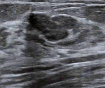
milla1cf Fri, 09/08/2023 - 16:05 September 8, 2023 — When diagnosing and treating cancer patients, the dynamics of blood flow to and from a suspected lesion can provide clinicians with valuable diagnostic information. Visit Philips EPIQ Elite for more information on Philips premium ultrasound.
Diagnostic imaging is an important tool used every day in healthcare to assist doctors in making the most informed decisions for their patients. Over the years, advancements in diagnostic imaging have greatly increased patients’ overall care, quality of life, and outcome when diagnosed with certain conditions.
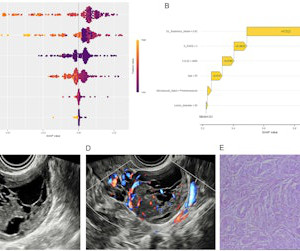
An ensemble AI model combining clinical variables, O-RADS, and deep learning radiomics can help in diagnosing ovarian tumors, suggest findings published March 5 in Translational Oncology. Radiology researchers continue to explore the potential of deep learning radiomics in more accurately diagnosing diseases through tumor differentiation.
Automated breast ultrasound (ABUS) with remote reading may improve access to breast cancer screening and early detection in low-resource settings, suggest findings published October 23 in the American Journal of Roentgenology. The team returned diagnostic reports to the community centers and patients sought follow-up care at local hospitals.
milla1cf Thu, 03/21/2024 - 10:42 March 21, 2024 — Artificial intelligence can spot COVID-19 in lung ultrasound images much like facial recognition software can spot a face in a crowd, new research shows. How does AI analyze ultrasound lung images?
Researchers have shown that an automated cancer diagnostic method, which pairs cutting-edge ultrasound techniques with artificial intelligence, can accurately diagnose thyroid cancer, of which there are more than 40,000 new cases every year.
Our results confirm the good diagnostic performance of F-18 FCH-PET/CT as the first-line functional imaging method for the detection of pathological parathyroid glands in patients with [primary hyperparathyroidism],” the group wrote. The study was published October 24 in Academic Radiology. The only curative treatment is to remove the glands.
S3A-SPBR-7 | Learning Center Compared with ultrasound or MRI used alone to diagnose breast diseases, a combined model of both modalities based on radiomics more accurately identifies breast conditions, according to a poster to be displayed Sunday morning. Sunday, December 1 | 11:45 a.m.-12:15 12:15 p.m. |
Combining automated breast ultrasound (ABUS) with contrast-enhanced ultrasoun. Read more on AuntMinnie.com Related Reading: Is axillary scanning needed in diagnostic breast ultrasound? Could ultrasound be used before DBT in focal breast complaints?
An artificial intelligence (AI) model using breast ultrasound images is on pa. Read more on AuntMinnie.com Related Reading: Is axillary scanning needed in diagnostic breast ultrasound? Systemic factors linked to incomplete breast imaging follow-up Could ultrasound be used before DBT in focal breast complaints?
We organize all of the trending information in your field so you don't have to. Join 5,000 users and stay up to date on the latest articles your peers are reading.
You know about us, now we want to get to know you!
Let's personalize your content
Let's get even more personalized
We recognize your account from another site in our network, please click 'Send Email' below to continue with verifying your account and setting a password.


Let's personalize your content