This site uses cookies to improve your experience. To help us insure we adhere to various privacy regulations, please select your country/region of residence. If you do not select a country, we will assume you are from the United States. Select your Cookie Settings or view our Privacy Policy and Terms of Use.
Cookie Settings
Cookies and similar technologies are used on this website for proper function of the website, for tracking performance analytics and for marketing purposes. We and some of our third-party providers may use cookie data for various purposes. Please review the cookie settings below and choose your preference.
Used for the proper function of the website
Used for monitoring website traffic and interactions
Cookie Settings
Cookies and similar technologies are used on this website for proper function of the website, for tracking performance analytics and for marketing purposes. We and some of our third-party providers may use cookie data for various purposes. Please review the cookie settings below and choose your preference.
Strictly Necessary: Used for the proper function of the website
Performance/Analytics: Used for monitoring website traffic and interactions
55881 Ablation of prostate tissue, transurethral, using thermal ultrasound, including magneticresonanceimaging guidance for, and monitoring of, tissue ablation. PC-6.47 $525.63 $209.28 PC-14.56 $8,508.75 $470.97 ICD-10 is the 10th edition of this coding system.
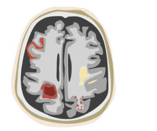
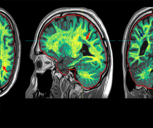
A large study, needed for FDA clearance, demonstrated that the use of icobrain aria significantly increases the accuracy of ARIA assessments by radiologists and hence allows for safer use of new amyloid-beta targeting therapies for Alzheimer’s disease patients.
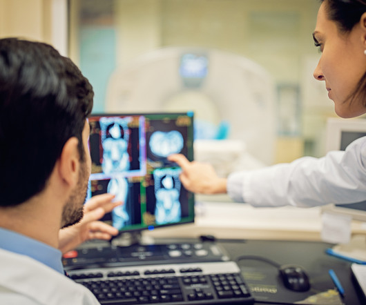
There are several types of diagnostic imaging available today; each one used to visualize the internal structures of the body to assist doctors in diagnosis and treating various diseases and medical conditions. Professional Radiology strives to provide patients with accurate and compassionate imaging services.
Computed tomography (CT scans) plays a critical role in diagnosing and monitoring a wide range of medical conditions. At Professional Radiology , we use state-of-the-art medical machinery to successfully provide patients in El Paso with accurate imaging services.
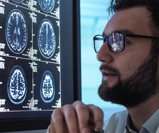
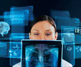
Integrating AI with our advanced imaging techniques will allow us to detect subtle changes in brain activity indicative of neurodegenerative diseases at their nascent stage.
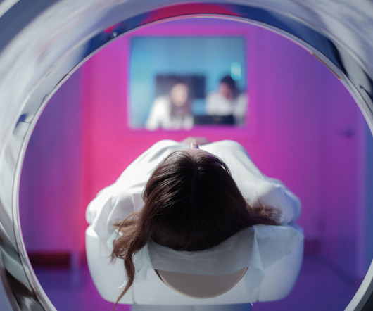
Since the advent of the magneticresonanceimaging (MRI) exam on human patients in the late 1970s, this innovation offered a multi-layered and noninvasive approach to the imaging of bodily organs, functions and the ability to diagnosedisease.
Computed tomography (CT scans) plays a critical role in diagnosing and monitoring a wide range of medical conditions. At Professional Radiology , we use state-of-the-art medical machinery to successfully provide patients in El Paso with accurate imaging services.
Diagnostic imaging is an important tool used every day in healthcare to assist doctors in making the most informed decisions for their patients. Over the years, advancements in diagnostic imaging have greatly increased patients’ overall care, quality of life, and outcome when diagnosed with certain conditions.
Magneticresonanceimaging (MRI) machines can clearly view non-bony parts of the body—soft tissue such as the brain, muscles and ligaments—as well as detect tumors, making it possible to diagnose many diseases and other conditions.
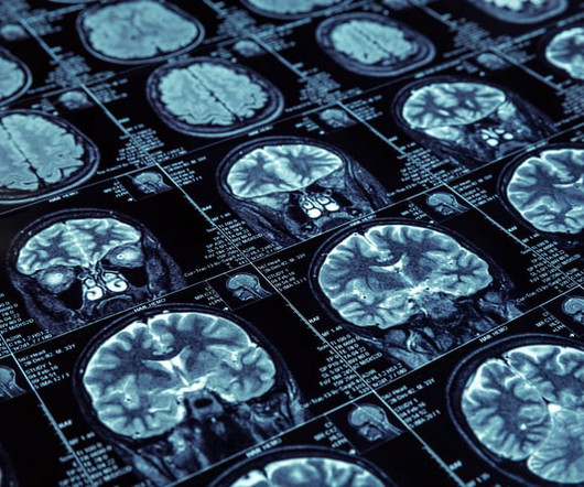
milla1cf Fri, 10/20/2023 - 18:29 October 20, 2023 — In certain cases, a new method can provide as much information from brain images taken with computed tomography (CT) as images captured with magneticresonanceimaging (MRI). About two percent of all people over the age of 65 are affected.
milla1cf Thu, 01/04/2024 - 10:47 January 4, 2024 — Diagnosing cancer today involves using chemical “contrast agents” to improve the accuracy of medical imaging processes such as X-rays as well as computed tomography (CT) and magneticresonanceimaging (MRI) scans.
Siemens Healthineers and UH will look to also advance the treatment of patients with Alzheimer’s disease and use theranostics—combining the approaches of diagnostics and therapeutics—to treat patients with advanced forms of certain cancers, as well as develop new magneticresonance ( MR ) technologies. tesla and 3T scanners.
milla1cf Thu, 07/06/2023 - 22:19 July 7, 2023 — Bayer , a global leader in radiology, has initiated the Phase III clinical development program called QUANTI , aiming to evaluate the safety and efficacy of gadoquatrane, an investigational extracellular macrocyclic gadolinium-based contrast agent (GBCA) for use in magneticresonanceimaging ( MRI ).
The world of medical imaging is marking a significant milestone in 2023: the 50th anniversary of magneticresonanceimaging (MRI). It assists in identifying diseases related to spine lesions, tumors, and stroke impacting the area of blood vessels and brain.
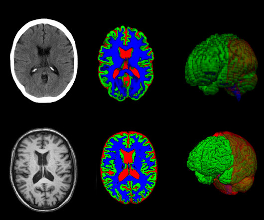
Accurate measurement of the volume and distribution of different tissue types in the brain is important for diagnosing brain disease. However, visual assessments of MR images are often subjective, and lack the necessary accuracy while measuring tissue volume from 2D image slices is virtually impossible.
milla1cf Fri, 03/29/2024 - 08:01 March 29, 2024 — Magneticresonanceimaging ( MRI ) is a cornerstone in the landscape of medical diagnostics, celebrated for its non-ionizing and non-invasive nature. For more information: [link] Friday, March 29, 2024 - 08:01
During the Radiological Society of America Scientific Sessions and Annual Meeting, RSNA 2023 , GE HealthCare showcased a number of AI and deep learning enabled solutions that help address these challenges, including the introduction of the new SIGNA Champion wide bore magneticresonanceimaging (MRI) system.
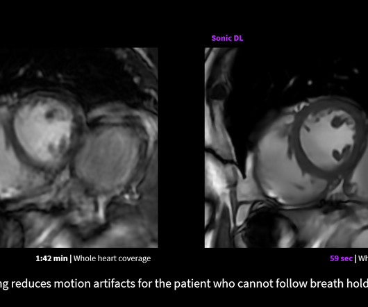
milla1cf Wed, 06/07/2023 - 19:51 June 6, 2023 — GE HealthCare announced the FDA clearance and launch of Sonic DL – a state-of-the-art deep learning-based technology designed to dramatically accelerate image acquisition in magneticresonanceimaging ( MRI ).
The imaging evaluation of chronic mesenteric ischemia (CMI) is usually first performed with a duplex ultrasound study. As seen with other disease patterns, many operators subsequently obtain cross-sectional or dynamic imaging for further evaluation.
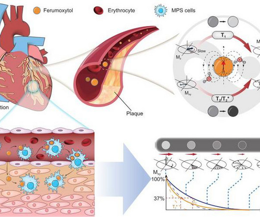
The state-of-the-art technology uses magneticresonanceimaging (MRI) to create detailed images of the heart. The team say their findings could help doctors better diagnose and monitor patients with heart disease and heart failure. New technology can detect rapid pressure changes inside your heart.
MagneticResonanceImaging (MRI) MRI stands for MagneticResonanceImaging. Instead, they employ a powerful magnetic field along with radio waves to produce detailed images of organs and tissues. Dental Assessment: Helps in diagnosing tooth decay and periodontal disease.
Magneticresonanceimaging (MRI) is used to help diagnose and treat various medical conditions. At Intermountain Medical Imaging , we rely on a variety of MRI options that offer a wider opening, helping us deliver high-quality care to clients and medical providers alike. MRIs rely on large magnets and radio waves.
According to the American Thyroid Association, an estimated 20 million Americans have some form of thyroid disease. However, up to 60% of those with thyroid disease are unaware of their condition. Blood tests Imaging tests Physical exams What testing can be used to diagnose thyroid disorders?
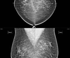
The approval expands upon Bayer's focus on breast imaging, with a portfolio that also includes Gadavist (gadobutrol) injection, a gadolinium-based contrast agent approved for use with MRI ( MagneticResonanceImaging ) to assess the presence and extent of malignant breast disease in adult patients.
Magneticresonanceimaging (MRI) is an essential tool that healthcare providers use to diagnose and treat various conditions. It provides highly detailed images of what is going on inside the body and how multiple systems work with each other.
Radiology and medical imaging are intertwined in medical diagnostics. Radiology is a branch of medicine that uses radiant energy in the diagnosis and treatment of disease. Practitioners of radiology are called radiologists, and they utilize imaging technology in the diagnosis and treatment of patients.
In 2011, a large study examined the use of x-rays and other radiation imaging on children—they estimated that the average child would get more than seven radiation scans by the age of 18. No doubt, then, that the role of a pediatric radiologist is important in accurately diagnosing and treating diseases and conditions in children.
Introduction: MagneticResonanceImaging (MRI) has transformed the landscape of modern healthcare, enabling us to see beyond what the eye can perceive. Chapter 1: Unveiling the Magic of MRI An introduction to the enchanting world of MagneticResonanceImaging.
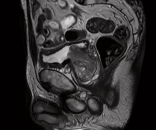
19, 2024 — GE HealthCare recently announced a collaboration with the University of California San Diego School of Medicine to investigate advanced magneticresonanceimaging (MRI) protocols and techniques for female-specific diseases and conditions of the pelvis and develop comprehensive educational materials for clinicians.
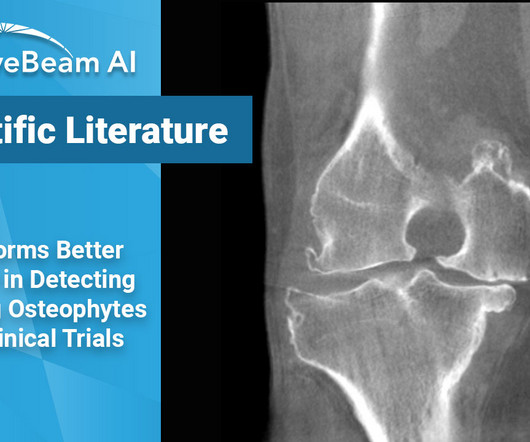
Key Points: While magneticresonanceimaging (MRI) adequately detects the size and presence of osteophytes (OPs) in the medial tibio-femoral compartment, it underestimates OPs in all other knee compartments of osteoarthritic patients. In the research setting, weight bearing CT (WBCT) imaging could be a useful tool.
As radiology departments proliferated in hospitals, X-rays became indispensable for diagnosing a wide range of conditions. They are used in the diagnosis of bone fractures, lung diseases, dental issues, and various other medical conditions. Advanced Imaging Modalities: X-ray technology has expanded beyond conventional radiography.
MRI-Scan-Teleradiology Introduction: MagneticResonanceImaging (MRI) has evolved significantly since its inception, and the latest breakthroughs in technology are transforming the field of medical imaging. Its significance in diagnosing vascular diseases and planning interventions.
Introduction: MagneticResonanceImaging (MRI) is often described as a wizardry of modern medicine, offering a glimpse into the human body’s inner workings with astonishing detail. Real-world tales of how MRI has been a wizard’s wand in diagnosing and treating medical conditions.
Among them, so-called spiral sequences that could reduce scanning time, e.g., when diagnosing blood clots, sclerosis and tumors. Spiral sequences would also be an attractive tool in MRI research, where, among other things, they could provide researchers and health professionals with new knowledge about brain diseases.
It is a medical imaging system used to store, manage, and share cardiology images. Cardiologists use CPACS systems to view, diagnose, and treat heart conditions. Like PACS , CPACS systems typically use DICOM (Digital Imaging and Communications in Medicine) standards to store and transmit images.
Doctors use imaging tests to see inside a patient’s body and diagnose their illnesses and injuries. There are many different types of imaging tests, each with their own method of generating a photo of the inside of the human body. A doctor will often use a diagnostic imaging test to check patients for signs of cancer.
This standard has revolutionized the radiology industry, encompassing many imaging modalities such as X-rays, computed tomography (CT), magneticresonanceimaging (MRI), ultrasound, nuclear medicine, PET scans, etc. Practicing Radiologists: access DICOM image files for studying, interpreting, and diagnosing them.
A CVIS (or CIS) is a specialized software platform designed to manage, store, and analyze clinical information related to diagnosing, treating, and managing cardiovascular diseases. In another example, consider the case of a patient with a history of heart disease who visits the hospital due to chest pain.
tesla MRI AI body composition analysis Cardiac PET Cryo/thermoablation CT colonography Genicular artery embolization Hyperpolarized xenon-129 MRI PET/MRI Photon-counting CT Radiomics Theranostics Whole-body MRI screening Image of the Year 3D PET/MR image. Image from Eric Guedj, MD, PhD, of Marseille University Hospital, et al.
Diagnostic imaging tests are tools used by physicians to diagnose a range of medical conditions. Each of these imaging methods uses different technologies to create real-time images and videos of the internal structures of the body. There is no need to be anxious as imaging tests are non-invasive and painless.
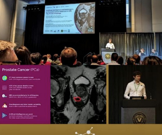
Prostate cancer is the most frequently diagnosed cancer in men and is a leading cause of death in men, second only to lung cancer. Understanding The Disease Types of prostate cancer range from low-grade to advanced, and call for different types of treatment plans. Prostate Cancer Awareness September is Prostate Cancer Awareness Month.
Medical imaging is used to help diagnose these injuries, so doctors can propose appropriate treatment plans. Diagnosing a Broken Bone Fractures almost always require emergency treatment. To determine if the bone is broken, doctors may use one of the following imaging technologies. What Are Broken Bones?
Currently, only 10% to 20% of patients are diagnosed at a stage where the tumor is resectable. Magneticresonanceimaging ( MRI ) plays an increasingly important role in the early diagnosis of prostate cancer. million new cases each year worldwide, accounts for 6.8% of male cancer-related deaths (350,000 deaths per year).
We organize all of the trending information in your field so you don't have to. Join 5,000 users and stay up to date on the latest articles your peers are reading.
You know about us, now we want to get to know you!
Let's personalize your content
Let's get even more personalized
We recognize your account from another site in our network, please click 'Send Email' below to continue with verifying your account and setting a password.


Let's personalize your content