This site uses cookies to improve your experience. To help us insure we adhere to various privacy regulations, please select your country/region of residence. If you do not select a country, we will assume you are from the United States. Select your Cookie Settings or view our Privacy Policy and Terms of Use.
Cookie Settings
Cookies and similar technologies are used on this website for proper function of the website, for tracking performance analytics and for marketing purposes. We and some of our third-party providers may use cookie data for various purposes. Please review the cookie settings below and choose your preference.
Used for the proper function of the website
Used for monitoring website traffic and interactions
Cookie Settings
Cookies and similar technologies are used on this website for proper function of the website, for tracking performance analytics and for marketing purposes. We and some of our third-party providers may use cookie data for various purposes. Please review the cookie settings below and choose your preference.
Strictly Necessary: Used for the proper function of the website
Performance/Analytics: Used for monitoring website traffic and interactions
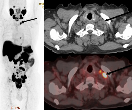
PET/CT scans with an experimental prostate-specific membrane antigen (PSMA) imaging agent can identify supraclavicular nodal metastasis in newly diagnosed prostate cancer patients, researchers have reported. For the analysis, the researchers included 240 patients who underwent scans for primary staging of newly diagnoseddisease.
The voters also zeroed in on the ongoing shortage of radiologists as the Biggest Threat to Radiology. Importantly, we are gaining deeper insights into disease processes themselves, which enhances our ability to diagnose and potentially treat conditions that were previously beyond our reach."
Multiparametric MRI (mpMRI) is a widely used approach for diagnosing patients with prostate cancer and reduces the need for invasive biopsies in approximately 30% of cases, Brembilla explained. The trial will eventually enroll 167 undiagnosed patients with suspected disease who have been referred for biopsies, Brembilla noted.
Technetium-99m (Tc-99m) methyl diphosphonate (MDP) bone scans are a potentially viable noninvasive option for diagnosing calciphylaxis, according to a team at the University of Massachusetts in Worcester, MA. All but one patient had renal disease and most had several other comorbidities, the authors wrote.
The findings were published February 21 in the Canadian Association of Radiologists Journal. The team assessed risk factors, disease stage, ICDR, biopsy rates, and positive predictive values for biopsy (PPV3). Gordon's group reported a positive predictive value for biopsy of 13% and an incidental cancer detection rate of 6.1
Our research is showing that we are missing opportunities to diagnosedisease early, Monticciolo told AuntMinnie.com. We need to have women, radiologists, and clinicians recognize these trends are not favorable and we have to reverse them if were going to save womens lives. We encourage women to get screened.
PET/MRI imaging shows promise in diagnosing fevers or inflammation of unknown origin and may have advantages over PET/CT, according to a study published January 3 in the European Journal of Radiology. Inflammation of unknown origin (IUO) is defined as prolonged and perplexing inflammation with temperatures below 38.3 °C.
The percentage of lung fibrosis quantified on CT pulmonary angiograms (CTPA) by an AI model is associated with increased risk of mortality -- and boosts clinicians' ability to predict the survival of lung disease patients, researchers have found. The AI model quantified fibrosis on the CT exams and was also scored by radiologists.
and radiology has learned much since then, according to experts who directly dealt with the diseases impact. While the pandemic affected medical operations across the country, the experts said that radiologists developed and honed their sense of resiliency as imaging was placed on the front lines.
A deep-learning model performs comparably to an abdominal radiologist when it comes to finding clinically significant prostate cancer on MRI, researchers have reported. External 0.86 External 0.86
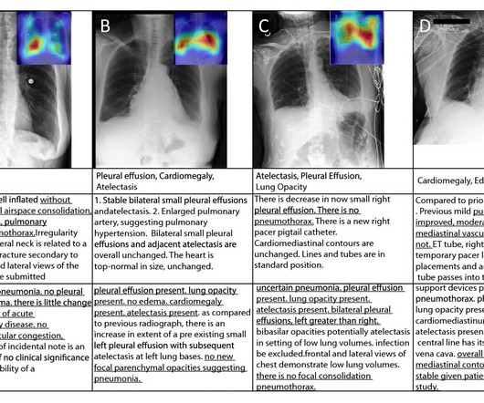
milla1cf Tue, 09/26/2023 - 15:32 September 26, 2023 — In a study of more than 2,000 chest X-rays , radiologists outperformed AI in accurately identifying the presence and absence of three common lung diseases, according to a study published in Radiology , a journal of the Radiological Society of North America ( RSNA ). Plesner, M.D.,
A large study, needed for FDA clearance, demonstrated that the use of icobrain aria significantly increases the accuracy of ARIA assessments by radiologists and hence allows for safer use of new amyloid-beta targeting therapies for Alzheimer’s disease patients. icobrain aria was thoroughly evaluated in large reader studies.
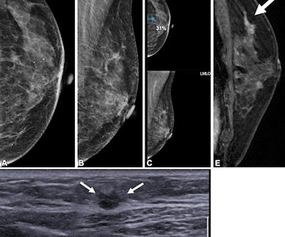
AI may have a role in finding contralateral disease in women with a personal history of breast cancer, according to findings published April 8 in Radiology. Lunit ) with that of two breast radiologists with no AI assistance for second breast cancer surveillance in women treated with unilateral mastectomy. Sensitivity 55.0%
milla1cf Wed, 08/02/2023 - 19:58 August 2, 2023 — An accepted manuscript published in the American Journal of Roentgenology (AJR) found that deploying a radiomic-based model with T2-weighted MRI data could increase diagnostic accuracy for pediatric Crohn disease (CD). Dillman et al.’s and accuracy of 89.6%. and accuracy of 89.6%.
A thyroid ultrasound imaging database could spur the development of more effective diagnostic and treatment models for related diseases, according to research published January 23 in Ultrasound in Medicine & Biology. Ultrasound is a first-line method in the screening and diagnosing of thyroid nodules.
Becoming a radiologist requires an extraordinary level of dedication, education, and training. Radiologists are medical doctors who specialize in interpreting imaging studies like X-rays, CT scans, MRIs, and ultrasounds to diagnose and guide treatment for various conditions.
At the RSNA meeting, attendees will hear animated discussion on the opportunistic use of CT imaging, including, for example, how it can provide information on a patient's bone mineral density, catch pancreatic ductal adenocarcinoma earlier, and predict the presence of coronary artery disease by measuring epicardial fat volume. 9:20 a.m. |
A team led by Jan Rudolph, MD, from University Hospital, LMU Munich in Germany found that a convolutional neural network (CNN)-based AI system focusing on chest x-rays improved the performance of nonradiologists in diagnosing several chest pathologies. “In
Interobserver agreement for assessing interstitial lung disease (ILD) using high-resolution CT (HRCT) is moderate, researchers have reported. Our results demonstrate that repeatable assessment of disease severity, extent, and progression is challenging even for expert radiologists," the group noted.
The responses were scored as correct, incorrect, or clinically misleading by two cardiologists and one radiologist, with categorization based on majority vote. Questions ranged from those on simple diagnostic subjects (“My doctor wants to run tests to diagnose coronary artery disease. What tests will be ordered?”)
OpenAI's GPT-4 AI model can utilize imaging reports to generate summaries of disease course in patients with complex glioblastoma, improving treatment planning and potentially even enhancing radiology workflows, according to research published July 23 in Radiology. No image data were transmitted to GPT-4.
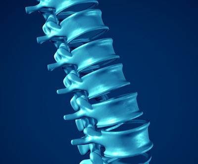
There are several types of diagnostic imaging available today; each one used to visualize the internal structures of the body to assist doctors in diagnosis and treating various diseases and medical conditions. It is often used to diagnose brain and spinal cord disorders, joint and musculoskeletal conditions, and cancer.
E16.3 - Other specified hypoglycemia E66.01 - Morbid (severe) obesity due to excess calories E66.09 - Other obesity due to excess calories Breast Cancer Biomarkers New Z codes indicate PR and HER-2 status, to be used alongside breast cancer diagnoses, facilitating tailored imaging protocols.
A novel machine-learning algorithm used with MRI can harmonize brain volumetric data of patients undergoing Alzheimer's disease assessment gathered from different scanners, researchers have reported. The findings were published December 18 in Radiology: Artificial Intelligence. The complete study can be found here.
The potential benefit of abbreviated MRI is reduced scan time for the patient, increased patient throughput, and decreased interpretation time for the radiologist," she told session attendees on November 26. response category 0.68 Detection of new lesions 0.49
Read more on AuntMinnie.com Related Reading: PCCT allows radiologists to 'see more with less' Researchers sing PCCT's praises Short MRI with DWI improves accuracy in diagnosing breast cancer Can synthetic MRI replace conventional scans for brain imaging? New MRI contrast agent tops product news in September
“Given the rising incidence and geographical/ethnic variability of breast cancer in young women, physician awareness is crucial for timely diagnoses and addressing the significant impact of this disease,” Sefidbakht and co-authors wrote. Previous studies indicate that women are being diagnosed with breast cancer at a younger age.
Integrating AI with our advanced imaging techniques will allow us to detect subtle changes in brain activity indicative of neurodegenerative diseases at their nascent stage.
When it comes to detecting and diagnosing various medical conditions, the field of radiology plays a critical role. Professional Radiology, a leading diagnostic imaging radiology center in El Paso, has been at the forefront of these innovations, offering a wide range of services to aid in the early detection and diagnosis of diseases.
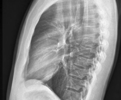
As I was on the phone with a colleague trying to convince the referrer of why I think a patient has Paget’s disease instead of metastases, I described the cortical thickening of the iliopectineal line and the lack of activity on the bone scan at the site and elsewhere throughout the body. Does this technique sound familiar?
CT-guided percutaneous needle biopsy is a minimally invasive procedure used to analyze tissue and is key in diagnosing suspicious pulmonary nodules, the authors explained. Patient with extensive lung and pleural disease. (2a) They then compared the accuracy of biopsies among the groups.
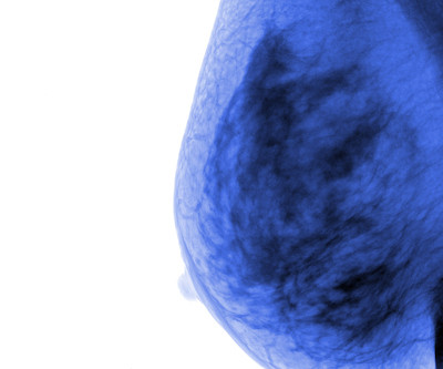
Breast cancer remains the most commonly diagnosed cancer among women in the U.S. i Thankfully, the field of breast screening is not static, and advancements are revolutionizing how we detect and diagnose the disease. Erik Anderson, president, breast & skeletal health solutions at Hologic.
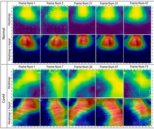
AI could become a crucial tool for radiologists, with recent advancements enabling it to accurately diagnose pneumonia, COVID-19, and other lung diseases.
"This collaborative multimodal approach not only boosts diagnostic performance but also offers interpretable insights to radiologists, paving the way for advanced, efficient disease diagnosis and improved clinical decision-making," the authors noted. The researchers extracted textual features (i.e., Microaveraged AUC 0.89
Alzheimer disease is a progressive, irreversible brain disorder that slowly degrades memory and cognitive function. While previous treatment methods focused on addressing Alzheimer disease symptoms, recent approvals of monoclonal antibodies have provided a path to target the underlying disease itself. In June 2021, the U.S.
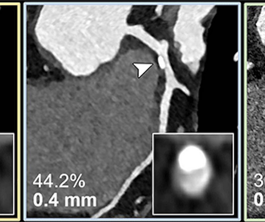
Coronary CT angiography is a first-line test in the assessment of coronary artery disease. attending radiologist at the University Medical Center Mainz in Germany, and assistant professor of radiology at the Medical University of South Carolina in Charleston.
AI software can come to the aid of overwhelmed radiologists by aiding in the crucial tasks of detection, quantification, and future risk prediction of lung cancer on CT exams -- both in low-dose screening exams and in nonscreening chest CT exams. We are diagnosing more nodules than ever before.
Considering a patient's symptoms as noted on a short pre-MR imaging questionnaire improves radiologists' lumbar spine exam interpretation and diagnosis, researchers have found. Their diagnoses were then compared to those of spine specialists using Cohen kappa values. The results were published October 29 in Radiology.
CNNs have been used in AI research to classify breast cancer disease status and predict therapeutic responses based on diagnostic images of the primary breast tumor. Finally, two expert radiologists isolated the regions of interest of the primary tumor from other areas of breast tissue.
In most cases, the radiologist merely has to confirm the suggested diagnosis—e.g., According to the creators of the artificial intelligence solution, the technology lowers this time to about 30 seconds when no considerable text revision is required. fibrosis, enlarged heart, or a suspected malignant tumor—or absence thereof.
The research included 746 patients from a longitudinal interstitial lung disease database; of these, 525 made up a low-risk ILA cohort and 221 made up a high-risk cohort. Schnitzler and colleagues conducted a study to identify imaging and clinical features on CT that could predict a patient's progression from ILA to IPF.
Such algorithms can help teach computers how to detect and diagnosedisease, the NIH said in a September 27 announcement. A number of the images are of patients with advanced lung disease, according to the NIH. The database is described in a paper published earlier this year on the website of the Computer Vision Foundation.
At TRA Medical Imaging, we are proud to offer low-dose lung cancer screenings (LDCT) to patients who meet specific criteria, providing a crucial tool in the fight against this disease. By the time many people are diagnosed, the cancer may have spread, making it harder to treat. Early detection can significantly change this trajectory.
We organize all of the trending information in your field so you don't have to. Join 5,000 users and stay up to date on the latest articles your peers are reading.
You know about us, now we want to get to know you!
Let's personalize your content
Let's get even more personalized
We recognize your account from another site in our network, please click 'Send Email' below to continue with verifying your account and setting a password.


Let's personalize your content