This site uses cookies to improve your experience. To help us insure we adhere to various privacy regulations, please select your country/region of residence. If you do not select a country, we will assume you are from the United States. Select your Cookie Settings or view our Privacy Policy and Terms of Use.
Cookie Settings
Cookies and similar technologies are used on this website for proper function of the website, for tracking performance analytics and for marketing purposes. We and some of our third-party providers may use cookie data for various purposes. Please review the cookie settings below and choose your preference.
Used for the proper function of the website
Used for monitoring website traffic and interactions
Cookie Settings
Cookies and similar technologies are used on this website for proper function of the website, for tracking performance analytics and for marketing purposes. We and some of our third-party providers may use cookie data for various purposes. Please review the cookie settings below and choose your preference.
Strictly Necessary: Used for the proper function of the website
Performance/Analytics: Used for monitoring website traffic and interactions
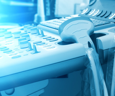
ChatGPT-4 demonstrated high accuracy in analyzing and interpreting thyroid and renal ultrasound images in a study published March 19 in Radiology Advances. These functionalities indicate the tool's potential to improve radiological workflows by pre-screening and categorizing ultrasound images,” Sultan told AuntMinnie.com.
ChatGPT-4 demonstrated high accuracy in analyzing and interpreting thyroid and renal ultrasound images in a small study published March 19 in Radiology Advances. These functionalities indicate the tool's potential to improve radiological workflows by pre-screening and categorizing ultrasound images,” Sultan told AuntMinnie.com.
For communities in Illinois and Ohio, where many rural hospitals and outpatient centers serve as lifelines, ensuring timely and accurate radiological services is crucial. By reducing overhead costs associated with maintaining in-house radiologists, rural hospitals can ensure uninterrupted imaging services without compromising care quality.
AI improves chest x-ray imaginginterpretation by nonradiologist practitioners, which could be useful in low-resource settings, according to research published January 29 in Chest. On internal validation, the AI algorithm achieved area under the curve (AUC) values ranging between 0.95 (nodules) and 0.995 (pleural effusion).
tesla MRI AI body composition analysis Cardiac PET Cryo/thermoablation CT colonography Genicular artery embolization Hyperpolarized xenon-129 MRI PET/MRI Photon-counting CT Radiomics Theranostics Whole-body MRI screening Image of the Year 3D PET/MR image.
A deep-learning method using endoscopic ultrasound images could detect pancreatic neuroendocrine neoplasms, according to research published October 17 in Gastrointestinal Endoscopy. Accuracy of CNN models, ultrasound users in detecting pancreatic neuroendocrine neoplasms Imaginginterpreter Accuracy CNN1 84.2% Expert user 85.5%
MNH at MUHAS is a public hospital serving Dar es Salaam, the largest city in Tanzania, and did not have a working mammography machine. For women in Tanzania, breast cancer is the second leading cause of cancer mortality,1 with more than 80% of diagnoses happening at stage III or IV when the odds of long-term survivability are much lower.2
Introduction: In Afghan hospitals, the implementation of teleradiology has been a game-changer, revolutionizing diagnostic capabilities and transforming healthcare delivery. Many patients in remote areas face hurdles when trying to access healthcare facilities, especially those equipped for diagnostic imaging.
Introduction: Teleradiology has emerged as a transformative solution for hospitals, providing a seamless way to enhance diagnostic capabilities and streamline radiology services. Extended Coverage and 24/7 Availability: Ensuring Timely Diagnoses: Explore how teleradiology extends coverage beyond traditional working hours.
In 2025, AI tools are more refined than ever, assisting radiologists with cancer detection, anomaly identification, and imageinterpretation. Advanced algorithms can now process vast amounts of imaging data faster than ever, reducing turnaround times and enhancing patient outcomes.
PET-CT-Scan-Reporting-Service Introduction: The integration of teleradiology services into hospital systems has emerged as a transformative advancement in modern healthcare. Discuss how teleradiology allows hospitals to tap into a pool of skilled radiologists irrespective of geographical locations, ensuring prompt and accurate diagnoses.
PET-CT-Scan-Reporting-Service Introduction: Teleradiology has emerged as a transformative force, extending healthcare services beyond the confines of traditional hospital settings. Remote ImageInterpretation: Explore how teleradiology enables remote imageinterpretation.
A few of the key findings in the survey report include: POCUS adoption has stalled due to a lack of infrastructure The top challenges caregivers face with their current POCUS solutions are poor image quality and integrating the device with hospital IT such as PACS or electronic health records (EHR).
Global Accessibility: Geographical constraints become obsolete as teleradiology enables remote interpretation of medical images. Rapid Diagnoses and Treatment: Teleradiology ensures swift and efficient diagnoses, particularly critical in emergency scenarios.
Accessibility Beyond Borders: Geographical barriers crumble as teleradiology enables remote interpretation of medical images. Rapid Diagnoses and Treatment: Teleradiology facilitates swift and efficient diagnoses, particularly crucial in emergency situations.
Example: Diagnosing a small atrial septal defect (ASD) in a transthoracic echocardiogram (TTE) can be challenging due to its subtle presentation and the need to differentiate it from normal anatomical variations. Identifying subtle anomalies like small congenital defects or early signs of disease can be difficult.
Finally, most hospitals lack the necessary protocols and processes to coordinate the complex interdisciplinary care patients with RAO may require. Diagnosed with a nonarteritic retinal artery occlusion (RAO) based on OCT findings and follow-up examination. Paper: Lema GMC, De Leacy R, Fara MG, et al. Ophthalmology.
Versatile Mobile Reading Stations: Discuss the features and versatility of the mobile diagnostic reading stations from Imaging Solutions. Highlight capabilities such as on-the-go imageinterpretation, real-time reporting, and seamless integration with existing radiology workflows.
Teleradiology & Radiology data for artificial intelligence (AI) Introduction Belize is known for its stunning natural beauty, rich culture, and warm hospitality. It’s like having expert radiologists right here in Belize, offering faster and more accurate diagnoses. Speed can mean the difference between life and death.
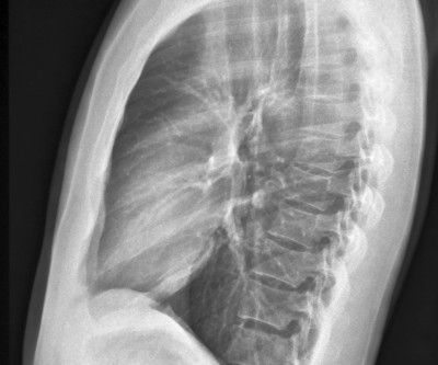
As radiology departments proliferated in hospitals, X-rays became indispensable for diagnosing a wide range of conditions. Artificial intelligence (AI) is also making its mark by enhancing imageinterpretation and diagnosis. Digital Transformation: The digital revolution transformed X-ray technology.
One of his tweets: #RGchat T1: the ABR certification exam is intended to test knowledge as it relates to competence, and critical thinking as it relates to imageinterpretation. The content will include critical findings as well as common and important diagnoses routinely encountered in general practice.
The Figure1 app on a mobile device This app is quick, easily accessible and helps radiologists, as well as other medical professionals, to get advice from peers across the world about diagnoses and treatment of various diseases and conditions.
This approach enables radiologists to focus on imageinterpretation rather than transcribing numbers, thereby reducing dictation time and minimizing errors. The study focused on the most common exams conducted at hospitals and imaging centers, including Complete Abdominal, Pelvic, and Thyroid exams. Nielson, J.,
Deep learning, a subset of machine learning, has significantly improved medical imaging analysis. Deep learning algorithms are trained to recognize specific markers in medical images, streamlining data analysis and improving diagnostic speed for accuracy.
This approach enables radiologists to focus on imageinterpretation rather than transcribing numbers, thereby reducing dictation time and minimizing errors. The study focused on the most common exams conducted at hospitals and imaging centers, including Complete Abdominal, Pelvic, and Thyroid exams. Nielson, J.,
Within weeks of his announcement hospitals world-wide had taken the initiative to open up X-ray rooms, which gave rise to the first radiology departments. (3) It was possible to obtain 3-D volumes when the images were taken at short intervals. (4) It was possible to obtain 3-D volumes when the images were taken at short intervals. (4)
We organize all of the trending information in your field so you don't have to. Join 5,000 users and stay up to date on the latest articles your peers are reading.
You know about us, now we want to get to know you!
Let's personalize your content
Let's get even more personalized
We recognize your account from another site in our network, please click 'Send Email' below to continue with verifying your account and setting a password.


Let's personalize your content