This site uses cookies to improve your experience. To help us insure we adhere to various privacy regulations, please select your country/region of residence. If you do not select a country, we will assume you are from the United States. Select your Cookie Settings or view our Privacy Policy and Terms of Use.
Cookie Settings
Cookies and similar technologies are used on this website for proper function of the website, for tracking performance analytics and for marketing purposes. We and some of our third-party providers may use cookie data for various purposes. Please review the cookie settings below and choose your preference.
Used for the proper function of the website
Used for monitoring website traffic and interactions
Cookie Settings
Cookies and similar technologies are used on this website for proper function of the website, for tracking performance analytics and for marketing purposes. We and some of our third-party providers may use cookie data for various purposes. Please review the cookie settings below and choose your preference.
Strictly Necessary: Used for the proper function of the website
Performance/Analytics: Used for monitoring website traffic and interactions
The COVID-19 pandemic prompted an increase in the rates of negative mammograms in both screening and diagnostic settings, a study published November 14 in the Journal of Radiology Nursing found. They also found a decrease in the proportion of negative diagnostic mammograms.
They also spearheaded the development of guidelines for the American College of Radiology (ACR), recognizing the role that imaging had in diagnosing COVID-19 in patients. The guidelines included best practices for imaging with CT versus chest x-ray in diagnosing COVID, as well as whether imaging is necessary at all in some cases.
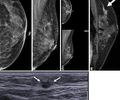
A team led by Su Min Ha, MD, PhD, from Seoul National University Hospital in South Korea reported that AI by itself achieved a higher performance than radiologists with no AI help when it came to detecting contralateral breast cancer in women treated with unilateral mastectomy. years after right mastectomy. (A) Sensitivity 55.0%
For communities in Illinois and Ohio, where many rural hospitals and outpatient centers serve as lifelines, ensuring timely and accurate radiological services is crucial. By reducing overhead costs associated with maintaining in-house radiologists, rural hospitals can ensure uninterrupted imaging services without compromising care quality.
A team led by Julie Hamzah, MBBS, from Singapore General Hospital, found that symptomatic first breast cancers, dense breasts, and the presence of trabecular thickening on mammography are tied to mammogram detection failure of ipsilateral second breast cancers.
The deployment aims to improve early detection of breast cancer, potentially reducing missed diagnoses and improving survival rates in Qatar's triennial screening program, according to the vendor. Two radiologists now read each mammogram while referring to Insight MMG's results, according to the firm.
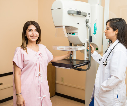
Breast cancer is the most commonly diagnosed form of cancer in American women. National initiatives are in place to urge women everywhere to get regular screenings, but there are some real disparities that exist when it comes to who is getting mammograms and how often.
The AI solution aims to enhance early detection of breast cancer, potentially reducing missed diagnoses and improving survival rates in Qatar's triennial screening program. Lunit's success in Qatar is part of a broader trend of the company's expansion in business-to-government (B2G) cancer screening initiatives worldwide.
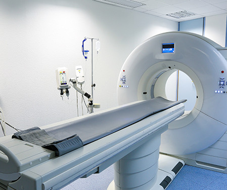
Shifts in Diagnostic Imaging: The Rise of Independent Facilities The trend of moving diagnostic imaging services away from hospitals and into Independent Diagnostic Testing Facilities (IDTFs) continues to grow in 2025. These facilities are adopting cutting-edge imaging technology, enabling faster and more accurate diagnoses.
For the study, 25 women, median age 52, recently diagnosed with breast cancer, underwent low-dose PEM with the radiotracer fluorine 18-labeled fluorodeoxyglucose ( 18 F-FDG). Two breast radiologists reviewed PEM images taken one and four hours post 18 F-FDG injection and correlated the findings with lab results. “It
The fact is, 75% of women diagnosed have no family history of breast cancer.If your family history is free of breast cancer, you should not neglect a yearly mammogram. Bottom line, if you are a woman over the age of 40, you should be getting a yearly mammogram. Myth: Mammograms can cause cancer.
At Manipal Hospitals Radiology Group, we believe in reaching out to the remotest corners and providing quality radiology reports which make a difference. Mutema’s vision, she entrusted the responsibility of making this care accessible to her patients to Manipal Hospitals Radiology Group (MHRG).
With an X-ray, Ultrasound, Mammogram and CT scan at our disposal, we needed to have a radiologist who could read all these modalities, and give us results in the shortest time possible to enable us to give the best medical care possible” she says. MHRG devised a 24/7 remote radiology solution for the Clinic.
It might be an MRI, an ultrasound, a mammogram, or another imaging test. When a primary care doctor orders specialized testing, say for a patient who complains of breast pain, they may not know the best imaging test to choose. Radiologists generally follow the American College of Radiology's Appropriateness Criteria to make these decisions.
All of the annual scheduled services such as mammograms can now be scheduled, as well as imaging prescribed by physicians for the care of their patients. Patients may also schedule mammograms directly at our facilities in these same locations. What this means for our community and region is important.
Hence, mammograms carried out anywhere can now be viewed by expert breast radiologists in any part of the world thanks to teleradiology. If the breast is dense on the mammogram, an ultrasound must also be carried out. most breast cancers diagnosed after age 50. Age 40-50 Once in 2 years 3. Getting older.-most
A) Mammogram MLO view. Mammogram CC view. A mammogram demonstrated focal asymmetries involving most of the anterior and mid right breast with diffuse skin thickening, trabecular coarsening, increased overall density, and enlarged right axillary lymph nodes. What is the diagnosis? Xray of the Week Figure 1.
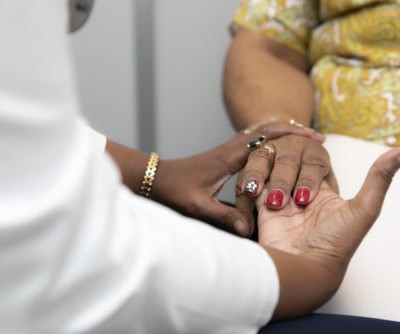
When it comes to accurate diagnoses and effective patient care, getting a second opinion on imaging results can make all the difference. Our abdominal imaging specialists ensure accurate diagnoses for conditions like pancreatic cancer or complex GI issues.
Doctors use imaging tests to see inside a patient’s body and diagnose their illnesses and injuries. This often involves blood, urine, and DNS tests, and medical imaging studies like mammograms, CT lung cancer screening scans and CT virtual colonography. Are Imaging Tests for Cancer only Available at Hospitals?
In addition, the ACR recommends that women diagnosed with breast cancer prior to age 50 or with a personal history of breast cancer and dense breasts should have annual supplemental breast MRI. Food and Drug Administration (FDA) requirement that all women having mammograms receive notice that their breasts are dense or not dense.
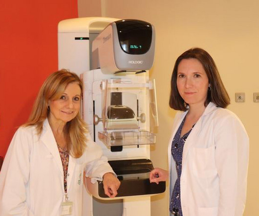
Breast cancer screening with mammography is well used, with 70% of eligible women reporting having undergone a mammogram within the past two years, Sandler noted. The study included 32,165 women who underwent screening mammography between November 2019 and December 2022 at two academic hospitals.
That is, it is diagnosed after a test whose result was negative and before the next evaluation. Conversely, if the number of tumors diagnosed after screening is low, it indicates that the program is fulfilling its function: to detect this disease at an early stage. per thousand people, while in the second it was 0.93.
However, Black women experience barriers to screening and are more likely to be diagnosed with triple-negative breast cancer at younger ages, and with advanced stage breast cancer. Only 31 percent had a documented mammogram in the EHR within the past two years. percent identified as Black/African American.
A study by researchers from the Cancer Center at West China Hospital of Sichuan University in China delved into this connection, utilizing genetic data from genome-wide association studies (GWAS). While some studies suggest a higher breast cancer risk in individuals with migraines, others indicate the opposite or mixed results.
You’re more likely to be diagnosed with breast cancer than any other cancer (besides skin cancer). That decline has been attributed, in large part, to annual screening mammograms. Each year, during your mammogram appointment, a full assessment is done to determine if you have one or multiple risk factors.
Within weeks of his announcement hospitals world-wide had taken the initiative to open up X-ray rooms, which gave rise to the first radiology departments. (3) Portrait of Sir Godfrey Hounsfield (1919-2004) The first clinical CT scan: Atkinson Morley's Hospital, October 1971 Credit: impactscan.org. Ok, so what do radiologists do now?
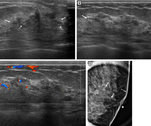
A team led by Su Min Ha, MD, PhD, from Seoul National University Hospital in South Korea found that the following nonmass lesion features on ultrasound can be considered suspicious: calcifications, posterior shadowing, segmental distribution, and mixed echogenicity. vs. 24% to 55%) than having a positive mammogram. cm vs. 1.9
We organize all of the trending information in your field so you don't have to. Join 5,000 users and stay up to date on the latest articles your peers are reading.
You know about us, now we want to get to know you!
Let's personalize your content
Let's get even more personalized
We recognize your account from another site in our network, please click 'Send Email' below to continue with verifying your account and setting a password.


Let's personalize your content