This site uses cookies to improve your experience. To help us insure we adhere to various privacy regulations, please select your country/region of residence. If you do not select a country, we will assume you are from the United States. Select your Cookie Settings or view our Privacy Policy and Terms of Use.
Cookie Settings
Cookies and similar technologies are used on this website for proper function of the website, for tracking performance analytics and for marketing purposes. We and some of our third-party providers may use cookie data for various purposes. Please review the cookie settings below and choose your preference.
Used for the proper function of the website
Used for monitoring website traffic and interactions
Cookie Settings
Cookies and similar technologies are used on this website for proper function of the website, for tracking performance analytics and for marketing purposes. We and some of our third-party providers may use cookie data for various purposes. Please review the cookie settings below and choose your preference.
Strictly Necessary: Used for the proper function of the website
Performance/Analytics: Used for monitoring website traffic and interactions
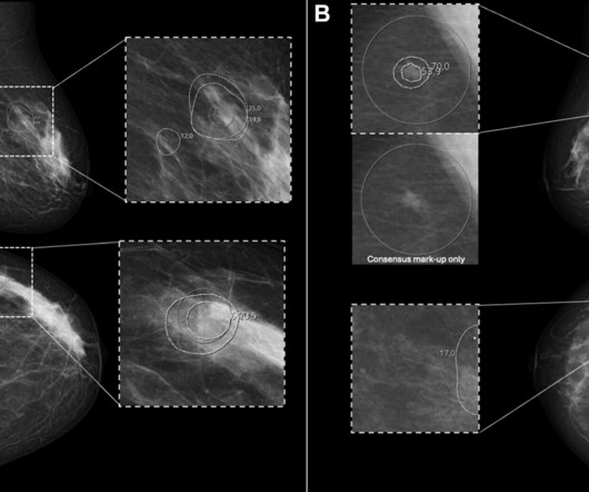
In a large-scale prospective trial conducted at a breast screening program in Europe, fewer women were recalled based on AI analysis of their digital mammogram than those recalled due to consensus findings from radiologists. The observed behavior may attenuate, and underestimate, the potential benefits of AI CAD in screening programs.
The voters also zeroed in on the ongoing shortage of radiologists as the Biggest Threat to Radiology. Importantly, we are gaining deeper insights into disease processes themselves, which enhances our ability to diagnose and potentially treat conditions that were previously beyond our reach."
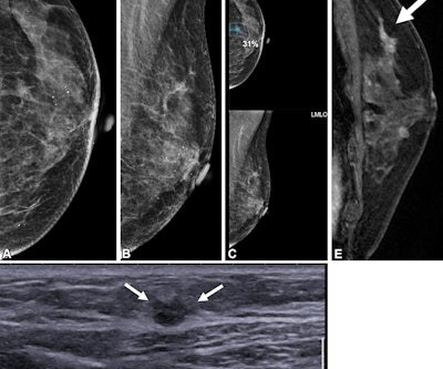
Women’s imaging at this year’s RSNA reflects momentous changes in trends regarding personalized, targeted healthcare in 2024 for women’s health. Research efforts toward supplemental breast imaging have ramped up in recent years and in Chicago, attendees can see results from these imaging modalities being put to the test.
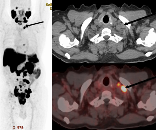
PET/CT scans with an experimental prostate-specific membrane antigen (PSMA) imaging agent can identify supraclavicular nodal metastasis in newly diagnosed prostate cancer patients, researchers have reported. For the analysis, the researchers included 240 patients who underwent scans for primary staging of newly diagnosed disease.
The finding is from a study that validated a fast protocol in 121 clinical patients, with seven independent readers giving the algorithm high scores for reducing image noise and improving image sharpness, noted lead author Jan Vosshenrich, MD, of New York University in New York City, and colleagues.
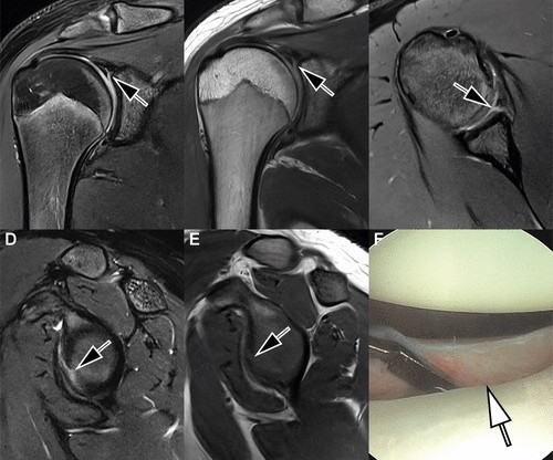
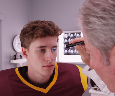
Sports radiologists are preparing for another fall season of high-contact sports, and that means brushing up on protocols and emphasizing teamwork among one's clinician colleagues. But getting ready for an uptick in musculoskeletal imaging doesn’t have to be daunting, according to Tim Klostermeier, MD, from UC Health in Cincinnati, OH.
With radiologists facing their fifth consecutive pay cut due to the 2025 Medicare Physician Fee Schedule, radiology practices are approaching hospital partners for stipends to continue offering their advanced services. Radiology professionals and groups must prepare now to adapt and thrive in this complex landscape.
Technetium-99m (Tc-99m) methyl diphosphonate (MDP) bone scans are a potentially viable noninvasive option for diagnosing calciphylaxis, according to a team at the University of Massachusetts in Worcester, MA. and Canada, which reported the sensitivity of bone scans for diagnosing calciphylaxis to be 62.5%, 89%, and 94.4%.
PET/MRI imaging shows promise in diagnosing fevers or inflammation of unknown origin and may have advantages over PET/CT, according to a study published January 3 in the European Journal of Radiology. T2 IDEAL WATER image (B) shows increased T2 signal; LAVA WATER (C) image shows mild enhancement in these muscles.
Multiparametric MRI (mpMRI) is a widely used approach for diagnosing patients with prostate cancer and reduces the need for invasive biopsies in approximately 30% of cases, Brembilla explained. Our preliminary data confirmed the potential increase of sensitivity of the combined use of PSMA PET with MRI compared to each modality alone.
In a study described as a “competition between radiologists,” participants tasked with identifying abnormal findings on chest x-rays performed better with AI assistance than without AI assistance – though not by much and not in all cases, according to a group in Nanjing, Jiangsu, China.
A deep-learning model performs comparably to an abdominal radiologist when it comes to finding clinically significant prostate cancer on MRI, researchers have reported. External 0.86 External 0.86
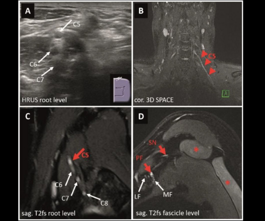
MRI and ultrasound have tradeoffs in diagnosing peripheral neuropathies of the upper extremity, a study published March 4 in Radiology found. This [MRN] may be the preferred imaging method when available, particularly for the suspected involvement of proximal nerve segments or multiple nerves, the Schwarz team wrote. Sensitivity 91.6%
While the pandemic affected medical operations across the country, the experts said that radiologists developed and honed their sense of resiliency as imaging was placed on the front lines. Medical imaging played a significant role in the early days of the pandemic when it hit its initial peak in April 2020.
ChatGPT-4 demonstrated high accuracy in analyzing and interpreting thyroid and renal ultrasound images in a small study published March 19 in Radiology Advances. These functionalities indicate the tool's potential to improve radiological workflows by pre-screening and categorizing ultrasound images,” Sultan told AuntMinnie.com.
when assessing text-based medical imaging cases, a study published January 16 in Radiology found. Large language models such as ChatGPT have interested radiologists within the past year for their ability to comprehend and generate human-like text. Imaging findings were originally characterized by radiologists.
ChatGPT-4 demonstrated high accuracy in analyzing and interpreting thyroid and renal ultrasound images in a study published March 19 in Radiology Advances. These functionalities indicate the tool's potential to improve radiological workflows by pre-screening and categorizing ultrasound images,” Sultan told AuntMinnie.com.
Virtual/augmented reality (VR/AR) headsets show promise for helping clinicians interpret CT exams -- even for conditions such as diverticulitis, which can be tricky to diagnose, researchers have reported. The results were published November 4 in the Journal of Imaging Informatics in Medicine. "[We to 5 for ease of use; from 3 to 4.7
Using direct radiologic image inputs can improve the diagnostic accuracy of large language models, according to research published July 9 in Radiology. The researchers highlighted that while previous studies have analyzed the performance of these models in radiologic settings, they used text-based inputs without images.
AI improves chest x-ray imaging interpretation by nonradiologist practitioners, which could be useful in low-resource settings, according to research published January 29 in Chest. That last part can be challenging for nonradiologists who do not constantly interpret diagnostic imaging exams.
Women in racial and ethnic minority backgrounds are less likely to be provided same-day diagnostic breast imaging services, despite such services being available, according to research published February 18 in Radiology. Additional imaging and possibly image-guided biopsy are recommended for women who have an abnormal screening mammogram. "For
Diagnostic Imaging is a great tool for your medical professional to use to detect issues sooner rather than later. There are several types of diagnostic imaging available today; each one used to visualize the internal structures of the body to assist doctors in diagnosis and treating various diseases and medical conditions.
This goes for women in all age groups and women who are Asian, Black, Hispanic, and Native American, according to the report written by Edward Hendrick, PhD, from the University of Colorado in Aurora and Debra Monticciolo, MD, from the Foundation for Imaging Research and Education in Temple, TX.
Imaging features are tied to pathological findings in breast cancer among young women, a study published January 26 in Clinical Imaging found. Previous studies indicate that women are being diagnosed with breast cancer at a younger age. Screening mammography is not typically recommended for women younger than 40 years of age.
ChatGPT would require further refinement before being used clinically to help educate patients about cardiac imaging, according to a study published May 23 in Clinical Imaging. The responses were scored as correct, incorrect, or clinically misleading by two cardiologists and one radiologist, with categorization based on majority vote.
The new Magnetic Resonance (MR) Safety Implant/Foreign Body Procedures of the Radiology/Diagnostic Radiology (Diagnostic Imaging) subsection of the CPT code book contains the codes and the guidelines for reporting them. They do not include any physician work value, but they would be available in the imaging center using global billing.
12, 2025 Konica Minolta Healthcare Americas, has published a case study by clinicians in the pulmonary and radiology departments at ASST Fatebenefratelli Sacco (Milan, Italy) demonstrating the use of Dynamic Digital Radiography (DDR) to help definitively diagnose diaphragm dysfunction. tim.hodson Fri, 02/14/2025 - 15:14 Feb.12,
Women’s imaging has a lot going for it at RSNA 2023 in Chicago. Supplemental screening takes center stage at the meeting as researchers highlight the roles of imaging modalities such as MRI, ultrasound, and molecular breast imaging in confirming suspicious findings on screening mammography.
Becoming a radiologist requires an extraordinary level of dedication, education, and training. Radiologists are medical doctors who specialize in interpreting imaging studies like X-rays, CT scans, MRIs, and ultrasounds to diagnose and guide treatment for various conditions.
Chest x-ray is used to diagnose RDS, but LUS has emerged as a reliable bedside tool for monitoring respiratory conditions in preterm infants. Images available for publishing under a creative commons license, CC BY-NC-ND 4.0. The NICU team was blinded to LUS findings while the radiologist was blinded to the x-ray chest report.
A team led by Su Min Ha, MD, PhD, from Seoul National University Hospital in South Korea reported that AI by itself achieved a higher performance than radiologists with no AI help when it came to detecting contralateral breast cancer in women treated with unilateral mastectomy. The radiologists had 10 and 17 years of experience, respectively.
Radiation dose and image quality performance measures for CT imaging accepted by the U.S. The new quality measure was adopted by the CMSin an effort to discourage excessive radiation dose while preserving image quality. Mahadevappa Mahesh, PhD,warns of potential "unintended consequences" of the CT measure.
Our results demonstrate that repeatable assessment of disease severity, extent, and progression is challenging even for expert radiologists," the group noted. High-resolution CT is a key imaging modality for evaluating ILD, the investigators explained, noting that "accurate classification of disease has important implications for patients."
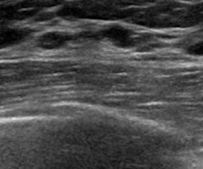
Sonographic imaging descriptors and histopathological diagnoses are needed for more accurate assessment of benign breast lesions, suggest findings published February 3 in BMC Womens Health. Still, this system is imperfect, with imaging findings of some benign breast lesions mimicking malignant characteristics.
Automation represents the current value of AI in the hospital emergency department (ED), a Yale University emergency radiologist shared last week at the 124th American Roentgen Ray Society (ARRS) meeting in Boston. We diagnose almost 180,000 pathologies as radiologists. It's usually a year-long process for budgeting."
A standardized detection reporting system can help radiologists accurately categorize how breast cancer is identified -- either through screening or symptomatic presentation -- when performing image-guided breast biopsies, researchers have found. These included the following: symptomatic presentations, imaging modalities, and other.
Breast cancer remains the most commonly diagnosed cancer among women in the U.S. i Thankfully, the field of breast screening is not static, and advancements are revolutionizing how we detect and diagnose the disease. Ultrasound rounds out the radiologist’s toolkit for supplemental imaging of women with dense breasts.
A thyroid ultrasound imaging database could spur the development of more effective diagnostic and treatment models for related diseases, according to research published January 23 in Ultrasound in Medicine & Biology. It also introduced a marker mask inpainting (MMI) method to erase artificial markers and improve image quality.
When it comes to your health, selecting the right diagnostic imaging center is an important decision. Whether you’re getting a routine scan or investigating a specific health concern, the imaging center you choose can significantly impact the accuracy of your diagnosis and the quality of care you receive.
Advanced imaging technologies continue to play a crucial role in detecting cancers before they progress, giving patients the best chance for successful treatment. The Importance of Early Detection Through Imaging Early detection of cancer through imaging allows for interventions at stages when treatment is most effective.
Food and Drug Administration (FDA) granted icometrix clearance for icobrain aria, the first AI software approved for detecting, measuring and grading amyloid-related imaging abnormalities (ARIA), a potentially harmful side effect of new amyloid-targeting therapies. icobrain aria was thoroughly evaluated in large reader studies.
Imaging and clinical features of interstitial lung abnormalities (ILA) can predict progression to idiopathic pulmonary fibrosis (IPF), according to research presented February 26 at ECR in Vienna. The investigators suggested that "future research should validate these findings in larger, multicenter cohorts."
We organize all of the trending information in your field so you don't have to. Join 5,000 users and stay up to date on the latest articles your peers are reading.
You know about us, now we want to get to know you!
Let's personalize your content
Let's get even more personalized
We recognize your account from another site in our network, please click 'Send Email' below to continue with verifying your account and setting a password.


Let's personalize your content