This site uses cookies to improve your experience. To help us insure we adhere to various privacy regulations, please select your country/region of residence. If you do not select a country, we will assume you are from the United States. Select your Cookie Settings or view our Privacy Policy and Terms of Use.
Cookie Settings
Cookies and similar technologies are used on this website for proper function of the website, for tracking performance analytics and for marketing purposes. We and some of our third-party providers may use cookie data for various purposes. Please review the cookie settings below and choose your preference.
Used for the proper function of the website
Used for monitoring website traffic and interactions
Cookie Settings
Cookies and similar technologies are used on this website for proper function of the website, for tracking performance analytics and for marketing purposes. We and some of our third-party providers may use cookie data for various purposes. Please review the cookie settings below and choose your preference.
Strictly Necessary: Used for the proper function of the website
Performance/Analytics: Used for monitoring website traffic and interactions
Women’s imaging at this year’s RSNA reflects momentous changes in trends regarding personalized, targeted healthcare in 2024 for women’s health. Research efforts toward supplemental breast imaging have ramped up in recent years and in Chicago, attendees can see results from these imaging modalities being put to the test.
Radiology is undergoing significant changes in 2025, driven by healthcare advancements, regulatory challenges, and workforce dynamics. Key trends include hospital consolidation of radiology services, the need for stronger cybersecurity, and innovative strategies to address staffing shortages.
Although relatively few of these changes will impact radiology practices, its essential to know what they are and adjust your practice systems accordingly. They do not include any physician work value, but they would be available in the imaging center using global billing. PC-6.47 $525.63 $209.28 PC-14.56 $8,508.75 $470.97
Microflow imaging can differentiate between benign and malignant breast lesions, suggest findings published October 28 in Clinical Radiology. Microflow imaging and high-definition microflow imaging can detect more blood flow in breast lesions than color Doppler flow imaging…,” Luo and co-authors wrote. Grade 1 25.4%
and radiology has learned much since then, according to experts who directly dealt with the diseases impact. While the pandemic affected medical operations across the country, the experts said that radiologists developed and honed their sense of resiliency as imaging was placed on the front lines.
Renaissance Imaging Medical Associates, a California-based Rad Partners affiliate, allegedly created false radiology reports supporting the diagnosis of spinal enthesopathy.
Food and Drug Administration (FDA) that use AI and machine learning, including those used in radiology. This legislation would create that system, improving diagnoses and encouraging the adoption of AI devices in clinical settings. cleared by the FDA are used in radiology. The Health Tech Investment Act (S. However, the U.S.
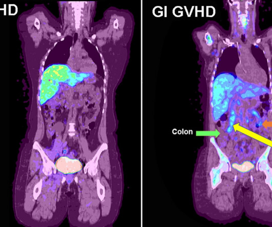
F-18 fluorothymidine (FLT) PET can identify early acute gastrointestinal graft versus host disease (GVHD) after patients undergo bone marrow transplants, according to a study published December 13 in Radiology: Imaging Cancer. F-18 FLT-PET imaging was performed on day 28. F-18 FLT-PET imaging of gastrointestinal GVHD.
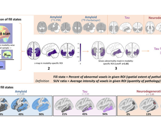
Researchers in Germany have proposed a new approach derived from brain PET imaging for diagnosing and staging Alzheimers disease, according to a study published March 25 in Radiology. Image and caption courtesy of RSNA. Schematic illustrates how fill states are derived. Con = control. The full study is available here.
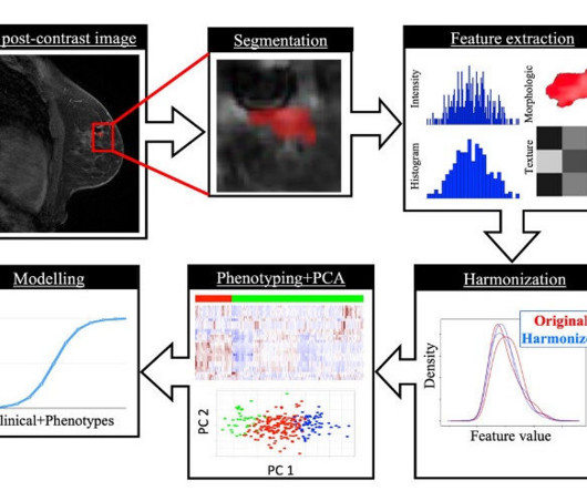
An MRI-based radiomics model shows potential for distinguishing low- from high-risk cases of ductal carcinoma in situ (DCIS), an early form of breast cancer, according to a study published April 1 in Radiology. Image courtesy of RSNA. About 20% of new breast cancers in the U.S. A graphical abstract of the study.
F-18 fibroblast activation protein inhibitor (FAPI) PET/CT leads to significant upstaging in newly diagnosed breast cancer, according to research published September 5 in Academic Radiology. The team put F-18 FAPI PET/CT to the test, comparing results to those of F-18 FDG PET/CT in systemic staging of newly diagnosed breast cancer.
PET/MRI imaging shows promise in diagnosing fevers or inflammation of unknown origin and may have advantages over PET/CT, according to a study published January 3 in the European Journal of Radiology. T2 IDEAL WATER image (B) shows increased T2 signal; LAVA WATER (C) image shows mild enhancement in these muscles.
FDG-PET imaging shows promise for use as a diagnostic criterion for neurosarcoidosis, with a recent case series illustrating the approach was effective when gold-standard approaches were not, according to a group of researchers in Berlin. “To Image courtesy of Neurological Research and Practice.
With 100% of precincts now reporting, we’re finally ready to declare this year’s winners in our annual awards program recognizing excellence in radiology. The voters also zeroed in on the ongoing shortage of radiologists as the Biggest Threat to Radiology. Most Influential Radiology Researcher Minnies 2024 Winner: Erik H.
F-18 FAPI-PET/CT is superior to F-18 FDG-PET/CT for diagnosing and staging patients with pancreatic cancer, according to a study published January 4 in the Journal of Nuclear Medicine. Typical PET (top), PET/CT (middle), and CT and MR (bottom) images of primary tumor obtained using both radiotracers in representative patients (A and B).
ChatGPT-4 demonstrated high accuracy in analyzing and interpreting thyroid and renal ultrasound images in a study published March 19 in Radiology Advances. These functionalities indicate the tool's potential to improve radiological workflows by pre-screening and categorizing ultrasound images,” Sultan told AuntMinnie.com.
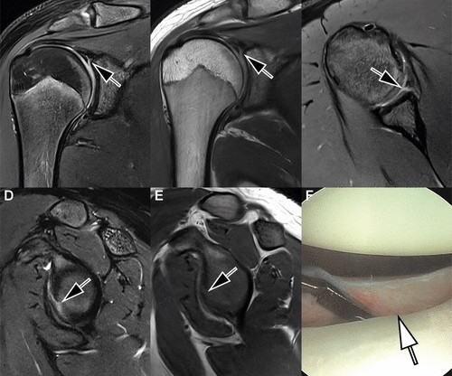
A commercially available deep-learning (DL) algorithm can enable good quality seven-minute shoulder MRI exams, according to research published February 18 in Radiology. According to the results, motion artifacts and image noise were minimal, with a ratings of 4 for each, and reconstruction artifacts were absent (a rating of 5).
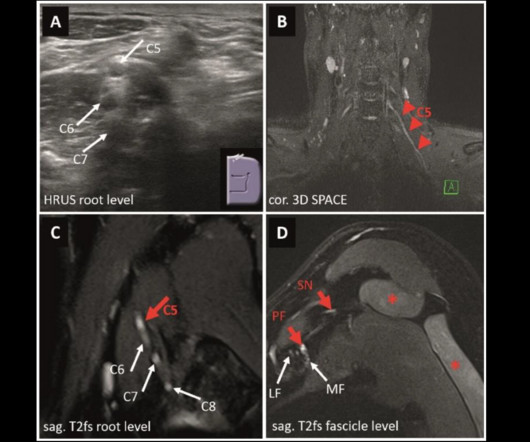
MRI and ultrasound have tradeoffs in diagnosing peripheral neuropathies of the upper extremity, a study published March 4 in Radiology found. This [MRN] may be the preferred imaging method when available, particularly for the suspected involvement of proximal nerve segments or multiple nerves, the Schwarz team wrote.
ChatGPT-4 demonstrated high accuracy in analyzing and interpreting thyroid and renal ultrasound images in a small study published March 19 in Radiology Advances. These functionalities indicate the tool's potential to improve radiological workflows by pre-screening and categorizing ultrasound images,” Sultan told AuntMinnie.com.
[This study] suggests a differential reliance on decision support related to whether that originated from AI CAD or from a fellow radiologist, the authors wrote in an article published March 18 in Radiology. All images and caption courtesy of the RSNA.
But getting ready for an uptick in musculoskeletal imaging doesn’t have to be daunting, according to Tim Klostermeier, MD, from UC Health in Cincinnati, OH. “I I love the role I play in diagnosing and treating these athletes. It [radiology] was a good fit for me because it’s kind of the architecture of the body. “It
when assessing text-based medical imaging cases, a study published January 16 in Radiology found. However, the researchers pointed out that it remains to be seen how upgrades in ChatGPT improve performance in providing diagnoses in radiology cases. and ChatGPT-4 in solving text-based Radiology “Diagnosis Please” cases.
Diagnostic Imaging is a great tool for your medical professional to use to detect issues sooner rather than later. There are several types of diagnostic imaging available today; each one used to visualize the internal structures of the body to assist doctors in diagnosis and treating various diseases and medical conditions.
Virtual/augmented reality (VR/AR) headsets show promise for helping clinicians interpret CT exams -- even for conditions such as diverticulitis, which can be tricky to diagnose, researchers have reported. The results were published November 4 in the Journal of Imaging Informatics in Medicine. "[We
As recently as three to five years ago, clinicians diagnosing disorders could not afford to dismiss the possible presence of “stumble-across” imaging studies. These are anatomic abnormalities that pop up in imaging exams looking for something else. Don’t confuse the term with incidental findings.
Augmented reality (AR) systems may enhance image-guided tumor ablations by improving the accuracy of needle placements, according to a study published January 29 in the Journal of Medical Imaging and Radiation Sciences. The full article is available here.
ChatGPT would require further refinement before being used clinically to help educate patients about cardiac imaging, according to a study published May 23 in Clinical Imaging. Questions ranged from those on simple diagnostic subjects (“My doctor wants to run tests to diagnose coronary artery disease.
Organizing pneumonia can hide underlying lung cancers in up to 10% of cases, and repeat imaging in the form of PET/CT -- and additional biopsy -- should be considered in patients with high clinical suspicion of malignancy, researchers have reported.
NPs purportedly would diagnose serious, complex conditions without the necessary medical imaging to reach such conclusions, the Department of Justice said Sept.
Using direct radiologicimage inputs can improve the diagnostic accuracy of large language models, according to research published July 9 in Radiology. The researchers highlighted that while previous studies have analyzed the performance of these models in radiologic settings, they used text-based inputs without images.
The trend may partially be attributable to radiologicdiagnoses becoming more common following “widespread deployment of sophisticated imaging technologies."
In a May 13 letter to representatives Kay Granger (R-TX) and Rosa DeLauro (D-CT) and senators Patty Murray (D-WA) and Susan Collins (R-ME), the groups noted that in radiology, AI is being used to help read and interpret images and help make more informed diagnoses.
AI improves chest x-ray imaging interpretation by nonradiologist practitioners, which could be useful in low-resource settings, according to research published January 29 in Chest. That last part can be challenging for nonradiologists who do not constantly interpret diagnostic imaging exams.
12, 2025 Konica Minolta Healthcare Americas, has published a case study by clinicians in the pulmonary and radiology departments at ASST Fatebenefratelli Sacco (Milan, Italy) demonstrating the use of Dynamic Digital Radiography (DDR) to help definitively diagnose diaphragm dysfunction. tim.hodson Fri, 02/14/2025 - 15:14 Feb.12,
POCUS allows for real-time evaluation and administration of treatment, without reliance on facility-based scanning, i.e. radiology departments, and avoidance of unnecessary tertiary care referral is a major benefit, he added. is a national platform for imaging AI, Tan explained. have created anatomical models based on medical images.
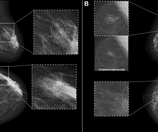
Women in racial and ethnic minority backgrounds are less likely to be provided same-day diagnostic breast imaging services, despite such services being available, according to research published February 18 in Radiology.
A generative adversarial network (GAN)-based noncontrast CT angiography (CTA) system has promise in vascular diagnosis, suggest research findings published November 14 in Radiology. Lyu and colleagues sought to develop and test its noncontrast deep learning GAN model to synthesize CTA-like images. and p > 0.99, respectively).
Although relatively few of these changes will impact radiology practices, it’s essential to know what they are and adjust your practice systems accordingly. Interventional radiology Dorsal Sacroiliac Joint Arthrodesis : New Category I code 27278 will replace Category III code 0775T, and existing code 27279 has been modified.
Sonographic imaging descriptors and histopathological diagnoses are needed for more accurate assessment of benign breast lesions, suggest findings published February 3 in BMC Womens Health. Still, this system is imperfect, with imaging findings of some benign breast lesions mimicking malignant characteristics.
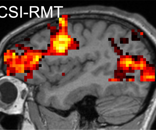
NYU Langone Health spinout Microstructure Imaging (MICSI) has secured U.S. Food and Drug Administration (FDA) 510(k) clearance for its MICSI-RMT AI image enhancement software for brain MRI. Designed to enhance white-matter imaging, MICSI-RMT employs a random matrix theory-based algorithm. Images courtesy of MICSI.
An advanced type of MR imaging shows that children and adolescents with long COVID have significant lung abnormalities, according to a study published February 25 in Radiology. Long COVID can affect people of all ages and is diagnosed when symptoms continue more than 12 weeks after the initial infection, the investigators noted.
CT imaging is best for diagnosing suspected intraorbital wooden foreign bodies, although MR imaging does offer valuable supplementary information, according to a study published February 20 in Clinical Radiology. Of the study participants, all 10 underwent CT imaging; five also underwent MR imaging.
We organize all of the trending information in your field so you don't have to. Join 5,000 users and stay up to date on the latest articles your peers are reading.
You know about us, now we want to get to know you!
Let's personalize your content
Let's get even more personalized
We recognize your account from another site in our network, please click 'Send Email' below to continue with verifying your account and setting a password.


Let's personalize your content