This site uses cookies to improve your experience. To help us insure we adhere to various privacy regulations, please select your country/region of residence. If you do not select a country, we will assume you are from the United States. Select your Cookie Settings or view our Privacy Policy and Terms of Use.
Cookie Settings
Cookies and similar technologies are used on this website for proper function of the website, for tracking performance analytics and for marketing purposes. We and some of our third-party providers may use cookie data for various purposes. Please review the cookie settings below and choose your preference.
Used for the proper function of the website
Used for monitoring website traffic and interactions
Cookie Settings
Cookies and similar technologies are used on this website for proper function of the website, for tracking performance analytics and for marketing purposes. We and some of our third-party providers may use cookie data for various purposes. Please review the cookie settings below and choose your preference.
Strictly Necessary: Used for the proper function of the website
Performance/Analytics: Used for monitoring website traffic and interactions
Image from Francisco Zamorano Mendieta, PhD, of the Universidad San Sebastián in Santiago, Chile Hypometabolism detected with F-18 FDG-PET in long-COVID patients : putative astrocyte dysfunction and glutamatergic dysregulation. Image from Eric Guedj, MD, PhD, of Marseille University Hospital, et al.
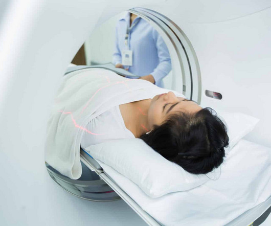
Diagnostic imaging tests are tools used by physicians to diagnose a range of medical conditions. Each of these imaging methods uses different technologies to create real-time images and videos of the internal structures of the body. There is no need to be anxious as imaging tests are non-invasive and painless.
The approval expands upon Bayer's focus on breast imaging, with a portfolio that also includes Gadavist (gadobutrol) injection, a gadolinium-based contrast agent approved for use with MRI ( MagneticResonanceImaging ) to assess the presence and extent of malignant breast disease in adult patients. for use in CEM.
Medical imaging is a technology which is used by radiologists , particularly for diagnostic purposes. Although the word “radiology” sounds like it involves radiation, that is not always the case – for example, MRI (magneticresonanceimaging) and ultrasound do not use radiation in their medical imaging technologies.
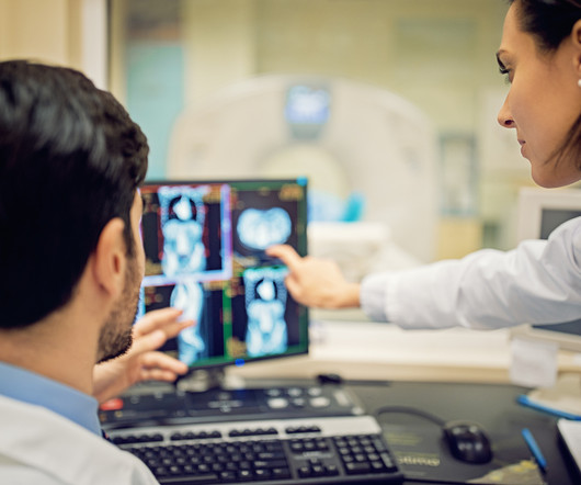
Radiologists and radiographers undertaking the imaging have most likely never seen the patient before. We need to bear in mind how we inform patients of the need for imaging and how it will help manage their symptoms. RadioGraphics. Journal of Clinical Nursing. 22(21-22):3225-3234. Patient-centred Radiology.
A) AP radiograph of Lisfranc Fracture Dislocation demonstrates the circled “fleck sign” or Lisfranc ligament avulsion fracture fragment. (B) C) The lateral radiograph notes with a circle, the dorsal sub dislocation of the metatarsal base. of all diagnosed fractures. Trauma due to falling off a roof. Xray of the Week Figure 1.
Computer aided diagnostic systems are already in use throughout radiology and can accurately diagnose breast cancer with a higher degree of accuracy than their human counterparts (2) and this use will only increase. 2011, Radiographics, pp. Legal aspects will need to be clarified if unsupervised AI reports are to start being issued.
after seeing the image. (2) Photoprint from radiograph by W.K. 3) In the early twentieth century, it was a common goal for investigators to try to find a way to separate the superimposed shadows that were recorded when a complex structure was shown on a radiograph. (3) 15) Radiologists needed a common means for sharing images.
Key Points: Imaging modalities such as plain radiographs (X-Ray), computed tomography (CT), and magneticresonanceimaging (MRI), dont have the diagnostic accuracy needed to detect syndesmotic widening or subtle instability. Specimens were mounted in a frame that allowed simulated axial weight bearing.
Magneticresonanceimaging and computed tomography in emergency assessment of patients with suspected acute stroke: a prospective comparison. Quant Imaging Med Surg. Comparative accuracy of CT perfusion in diagnosing acute ischemic stroke: A systematic review of 27 trials. Radiographics. Nadgir R, Yousef DM.
We organize all of the trending information in your field so you don't have to. Join 5,000 users and stay up to date on the latest articles your peers are reading.
You know about us, now we want to get to know you!
Let's personalize your content
Let's get even more personalized
We recognize your account from another site in our network, please click 'Send Email' below to continue with verifying your account and setting a password.


Let's personalize your content