This site uses cookies to improve your experience. To help us insure we adhere to various privacy regulations, please select your country/region of residence. If you do not select a country, we will assume you are from the United States. Select your Cookie Settings or view our Privacy Policy and Terms of Use.
Cookie Settings
Cookies and similar technologies are used on this website for proper function of the website, for tracking performance analytics and for marketing purposes. We and some of our third-party providers may use cookie data for various purposes. Please review the cookie settings below and choose your preference.
Used for the proper function of the website
Used for monitoring website traffic and interactions
Cookie Settings
Cookies and similar technologies are used on this website for proper function of the website, for tracking performance analytics and for marketing purposes. We and some of our third-party providers may use cookie data for various purposes. Please review the cookie settings below and choose your preference.
Strictly Necessary: Used for the proper function of the website
Performance/Analytics: Used for monitoring website traffic and interactions
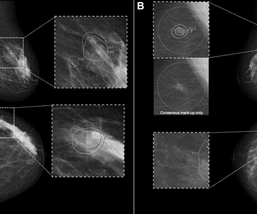
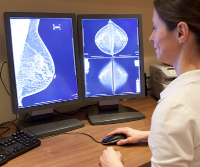
In a large-scale prospective trial conducted at a breast screening program in Europe, fewer women were recalled based on AI analysis of their digital mammogram than those recalled due to consensus findings from radiologists. Positive predictive value for breast cancer 3.4% 22% 25% 34.2% Positive predictive value for breast cancer 3.4%
The COVID-19 pandemic prompted an increase in the rates of negative mammograms in both screening and diagnostic settings, a study published November 14 in the Journal of Radiology Nursing found. They also found a decrease in the proportion of negative diagnostic mammograms.
Additional imaging and possibly image-guided biopsy are recommended for women who have an abnormal screening mammogram. The researchers identified multilevel factors associated with the availability of diagnostic services after an abnormal screening mammogram and women undergoing standard-of-care and advanced diagnostic services.
Our research is showing that we are missing opportunities to diagnose disease early, Monticciolo told AuntMinnie.com. Hendrick and Monticciolo said that many women unable to get their annual mammograms during the peak of the COVID-19 pandemic in 2020 have not returned to yearly screening. They added that the U.S.
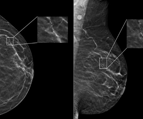
However, the researchers noted a lack of data on interpreting surveillance mammograms in women with a personal history of breast cancer. A) Left craniocaudal and (B) mediolateral oblique mammograms assessed as benign. (C) AI continues to show promise in improving screening mammography interpretation. years after right mastectomy. (A)
They also spearheaded the development of guidelines for the American College of Radiology (ACR), recognizing the role that imaging had in diagnosing COVID-19 in patients. The guidelines included best practices for imaging with CT versus chest x-ray in diagnosing COVID, as well as whether imaging is necessary at all in some cases.
From full-field digital mammograms, the AI extracted mammographic features including density, microcalcifications, masses, and left-right breast asymmetries for risk assessment. For these women, breast cancers were “more likely” diagnosed after two years follow-up, Eriksson said. The team also reported that 4.6% with 1 as reference).
Although a mammogram can not prevent cancer, it can help to diagnose cancer when it is treatable. In today’s post, we share some information that will hopefully put you at ease about having your first mammogram. How do you prepare for a screening mammogram? Ask us questions. Know before you go!
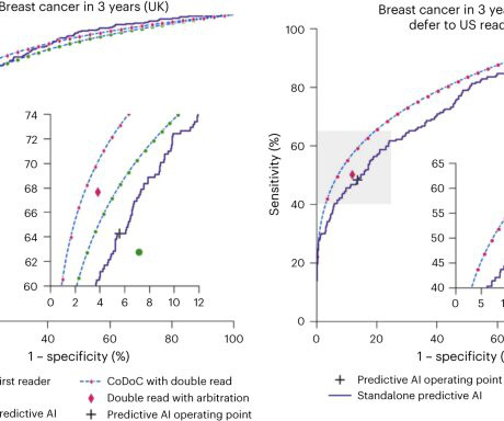
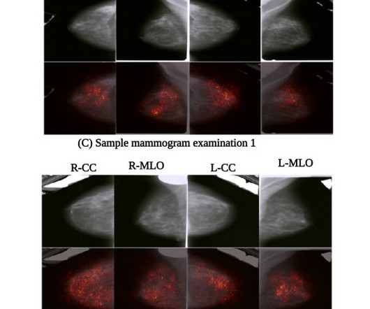
christine.book Tue, 05/21/2024 - 10:36 May 21, 2024 — According to a newly-published study of nearly 5,000 screening mammograms interpreted by an FDA-approved AI algorithm, patient characteristics such as race and age influenced false positive results. Nguyen, M.D.
When a mammogram reveals an abnormality or an individual is at a higher risk of breast cancer, diagnostic and supplemental imaging is required to determine if the patient needs a biopsy. Gina Curry (D-Delaware) and would eliminate costs for women for supplemental imaging such as breast MRIs and ultrasounds.
Breast cancer risk after a false-positive mammogram depends on individual patient characteristics and follow-up, a study published November 2 in JAMA Oncology found. Odds of developing breast cancer after false-positive mammogram Patient characteristic HR Women ages 60 to 75 2.02 Lower mammographic breast density 4.65 No biopsy 1.51
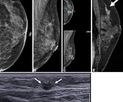
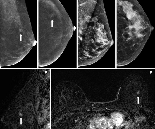
(B) Axial subtracted contrast-enhanced fat-suppressed T1-weighted image from a subsequent-round abbreviated MRI examination performed two years later shows a new 5-mm enhancing mass in the upper outer right breast (arrow), which was not seen on a mammogram performed five months prior. The exam was assessed as BI-RADS category 5. PPV2 21.3%
Gordon added that the guidelines could make way for more interval cancers diagnosed at advanced stages in younger women. If you’re in good health, keep having mammograms as long as you’re allowed to without a requisition, and then get a requisition,” she said.
A team led by Julie Hamzah, MBBS, from Singapore General Hospital, found that symptomatic first breast cancers, dense breasts, and the presence of trabecular thickening on mammography are tied to mammogram detection failure of ipsilateral second breast cancers.
ChatGPT-4 outperformed human clinicians in determining pretest and post-test disease probability after a negative test result involving chest radiographs and mammograms, according to a research letter published December 11 in JAMA Network Open.
“We're excited to introduce advanced workstation features for our flagship solution, ProFound Detection, aimed at further improving and facilitating radiologists' interpretation of mammograms within their workstation,” said Dana Brown , President and CEO of iCAD.
House Bill 2411 was introduced in the state by Representative David Cook (R-Globe) and includes eliminating costs for patients for MRI, ultrasound, and diagnostic mammograms. However, out-of-pocket costs for patients can range from $234 for a diagnostic mammogram to more than $1,000 for a breast MRI, according to the organization.
The reality is women are being diagnosed with heart attacks at an unprecedented rate,” Parghi said. “We We have a finding on a mammogram that can find women who can benefit from additional cardiovascular surveillance.” The team validated the algorithm’s accuracy on a dataset of 2D mammograms from 8,898 women.
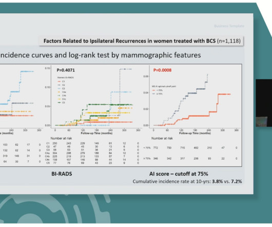
The researchers analyzed preoperative routine mammograms via a commercially available AI algorithm (Lunit Insight MMG, Lunit ). Yoon said that based on these results, more aggressive treatment may be considered in women diagnosed with DCIS showing AI scores at or greater than 75% on preoperative mammography.
It is estimated that 1 out of every 8 women in the United States will be diagnosed with breast cancer in her lifetime. One question our technologists are asked frequently is, “What’s the difference between a diagnostic mammogram and a screening mammogram?” first appeared on Clermont Radiology.
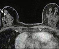
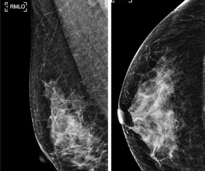
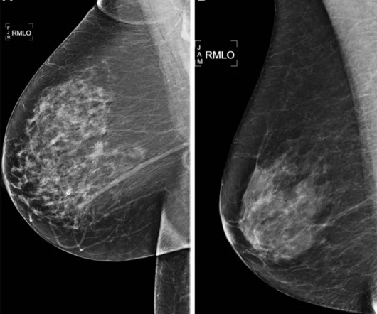
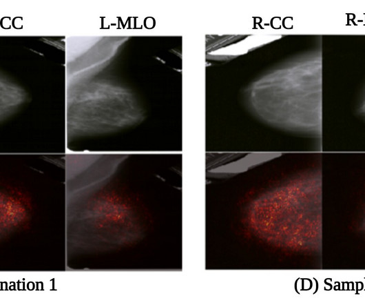
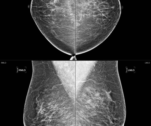
B) The right mediolateral oblique (RMLO) and (C) right craniocaudal (RCC) mammograms, obtained on the same day, show heterogeneously dense breast tissue that was assessed as being negative for cancer. (D) The research included 3,688 women who underwent breast cancer surgery between January 2009 and December 2014.
Response: While genetics can be a factor, 85% of women diagnosed with breast cancer have no family history. It is determined by a mammogram. The initiative addresses common misconceptions about the disease and about screening, including the following: Myth: I don’t have a family history of breast cancer, so I don’t need to worry.
The American Cancer Society recommends starting annual mammogram screenings at age 40. The list below are imaging exams used to diagnose breast cancer. Mammogram Screening Mammogram: Screening mammograms take 2 or more images of each breast. The list below are imaging exams used to diagnose breast cancer.
While such diagnosis technology can be used to help doctors diagnose breast cancer, the researchers also emphasized that “there is still much to be improved.” Sereshkeh's group explored the potential of a CNN-based deep learning model that incorporates deep features resulting from mammography staging. International.
The deployment aims to improve early detection of breast cancer, potentially reducing missed diagnoses and improving survival rates in Qatar's triennial screening program, according to the vendor. Two radiologists now read each mammogram while referring to Insight MMG's results, according to the firm.
A team of AI and medical specialists working with or for Google Research and Google DeepMind, has developed an AI based system designed to judge the confidence level of existing AI systems used for analyzing medical scans as a means of improving analysis of diagnostic tools, such as mammograms or chest X-rays.
A team led by Joao Horvat, MD, from the Memorial Sloan Kettering Cancer Center in New York found that CEM depicted 90% of breast cancers compared with 10% on low-energy mammograms alone and 50% on low-energymammogramswith whole-breast ultrasound. RSNA The team identified nine screen-detected cancers diagnosed in eight women.
A new artificial intelligence-based algorithm uses deep learning to analyze multiple mammogram views concurrently, simulating the evaluation process of radiologists.
milla1cf Fri, 06/09/2023 - 08:12 June 9, 2023 — In a large study of thousands of mammograms, artificial intelligence (AI) algorithms outperformed the standard clinical risk model for predicting the five-year risk for breast cancer. Something in mammograms allows us to track breast cancer risk. This is the ‘black box’ of AI.”
Breast cancer remains the most commonly diagnosed cancer among women in the U.S. i Thankfully, the field of breast screening is not static, and advancements are revolutionizing how we detect and diagnose the disease. Erik Anderson, president, breast & skeletal health solutions at Hologic.
Teleradiology: Bridging the Gap in Underserved Areas Teleradiology plays a pivotal role in expediting cancer diagnoses, particularly in rural or underserved regions. Investing in Advanced Imaging Technologies : Upgrading to the latest imaging equipment ensures higher resolution images, leading to more accurate diagnoses.
The algorithm provided a continuous cancer detection score for each exam ranging from zero to 100, with higher scores translating to a higher likelihood of cancer on the mammogram. Using exam data from between 2004 and 2018, the team evaluated the AI algorithm in 2022 and 2023. 79 Average differences (prior to interval cancers) 19.7
The earlier breast cancer is diagnosed, the easier it is to treat. Screenings for Breast Cancer The American Cancer Society estimates that more than 252,000 new cases of invasive breast cancer will be diagnosed in women in the U.S. To that end, you should be aware of the common signs and symptoms of breast cancer. this year alone.
Researchers at the University of Eastern Finland have developed a novel artificial intelligence-based algorithm, MV-DEFEAT, to improve mammogram density assessment. This development holds promise for transforming radiological practices by enabling more precise diagnoses. The study is published in IEEE Access.
Breast cancer is the most commonly diagnosed form of cancer in American women. National initiatives are in place to urge women everywhere to get regular screenings, but there are some real disparities that exist when it comes to who is getting mammograms and how often.
Another waited three months for mammogram findings. The Real-World Impact: Delayed Diagnoses, Frustrated Patients For hospitals and imaging centers, the shortage translates into longer turnaround times, heavier workloads, and sometimes critical delays. Waiting for answers that may change the course of their care.
Using mammography-based deep learning models may improve the accuracy of breast cancer risk assessment and can also lead to earlier diagnoses. “About 1 in 10 women develop breast cancer throughout their lifetime,” said study author Andreas D. Lauritzen , Ph.D., A variety of AI tools exist to aid in detecting cancer risk.
Radiologists must inform patients if they have dense breast tissue, a factor that can obscure mammogram results and increase cancer risks. Improving Patient Outcomes: Accurate and timely diagnoses lead to better treatment plans and improved patient care. Rural facilities are updating workflows to comply with these laws.
These facilities are adopting cutting-edge imaging technology, enabling faster and more accurate diagnoses. These laws require radiologists to inform patients if they have dense breast tissue, which can make it more difficult to detect cancer during mammograms.
Teleradiology-in-Flat-World Introduction : In the realm of modern medicine, diagnostic tools such as X-Rays, mammograms, and CAT scans play a crucial role in identifying and understanding various health conditions. Service Areas:- Rajasthan – Ajmer, Alwar, Banswara, Baran, Barmer, Bharatpur, Bhilwara, Bikaner, Bundi, Chittorgarh, Churu.
The AI solution aims to enhance early detection of breast cancer, potentially reducing missed diagnoses and improving survival rates in Qatar's triennial screening program. Lunit's success in Qatar is part of a broader trend of the company's expansion in business-to-government (B2G) cancer screening initiatives worldwide.
FACT: 90-95% of those diagnosed have no family history of breast cancer. FACT: 2,400 male breast cancer cases are diagnosed annually. MYTH: Mammograms can cause cancer to form or spread. FACT: Mammograms emit a very small dose of radiation, and the benefits of early detection far exceed any associated risks.
The data also suggests that 3D mammograms could reduce the incidence of advanced cancer diagnoses. Philpotts says fewer false positives and the overall higher cancer detection rate is a win-win and shows DBT is not over-diagnosing cancers. A randomized clinical trial to assess the impact of DBT is ongoing.
While fewer than 5% of women with breast cancer are diagnosed before the age of 40(1), those cancers are usually aggressive, and the young patients suffer from poor survival outcomes. Unfortunately, routine screening mammograms are not recommended for women under 40 because risks outweigh potential benefits at this young age.
We organize all of the trending information in your field so you don't have to. Join 5,000 users and stay up to date on the latest articles your peers are reading.
You know about us, now we want to get to know you!
Let's personalize your content
Let's get even more personalized
We recognize your account from another site in our network, please click 'Send Email' below to continue with verifying your account and setting a password.


Let's personalize your content