This site uses cookies to improve your experience. To help us insure we adhere to various privacy regulations, please select your country/region of residence. If you do not select a country, we will assume you are from the United States. Select your Cookie Settings or view our Privacy Policy and Terms of Use.
Cookie Settings
Cookies and similar technologies are used on this website for proper function of the website, for tracking performance analytics and for marketing purposes. We and some of our third-party providers may use cookie data for various purposes. Please review the cookie settings below and choose your preference.
Used for the proper function of the website
Used for monitoring website traffic and interactions
Cookie Settings
Cookies and similar technologies are used on this website for proper function of the website, for tracking performance analytics and for marketing purposes. We and some of our third-party providers may use cookie data for various purposes. Please review the cookie settings below and choose your preference.
Strictly Necessary: Used for the proper function of the website
Performance/Analytics: Used for monitoring website traffic and interactions
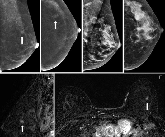
Abbreviated MRI may be suitable for screening women with dense breasts, a study published May 22 in the American Journal of Roentgenology found. Radiologists and ordering clinicians, alike, may use these results as evidence of the value of abbreviated MRI beyond the first round of screening in this population," Edmonds told AuntMinnie.com.
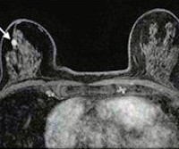
Postoperative MRI surveillance appears to lower the odds of advanced second breast cancer in women with a personal history of the disease, researchers have reported. "In Images in a 40-year-old woman who underwent breast-conserving surgery for left breast cancer and a surveillance breast MRI examination 25 months after surgery. (A)
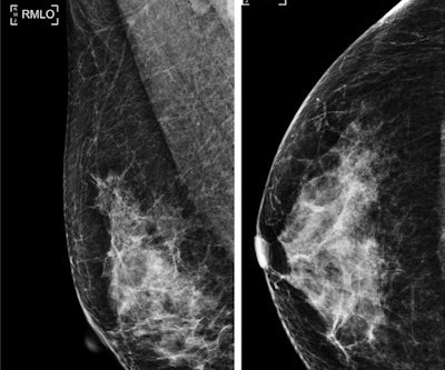
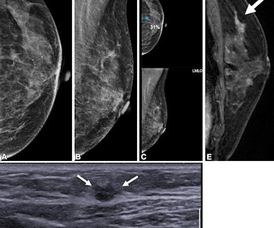
However, the researchers noted a lack of data on interpreting surveillance mammograms in women with a personal history of breast cancer. A) Left craniocaudal and (B) mediolateral oblique mammograms assessed as benign. (C) AI continues to show promise in improving screening mammography interpretation. years after right mastectomy. (A)
The USPSTF also said that there was insufficient evidence to recommend supplemental screening with MRI or ultrasound in women, regardless of breast density. Gordon added that the guidelines could make way for more interval cancers diagnosed at advanced stages in younger women.
House Bill 2411 was introduced in the state by Representative David Cook (R-Globe) and includes eliminating costs for patients for MRI, ultrasound, and diagnostic mammograms. However, out-of-pocket costs for patients can range from $234 for a diagnostic mammogram to more than $1,000 for a breast MRI, according to the organization.
A team led by Julie Hamzah, MBBS, from Singapore General Hospital, found that symptomatic first breast cancers, dense breasts, and the presence of trabecular thickening on mammography are tied to mammogram detection failure of ipsilateral second breast cancers.
While such diagnosis technology can be used to help doctors diagnose breast cancer, the researchers also emphasized that “there is still much to be improved.” Sereshkeh's group explored the potential of a CNN-based deep learning model that incorporates deep features resulting from mammography staging. International.
A team led by Joao Horvat, MD, from the Memorial Sloan Kettering Cancer Center in New York found that CEM depicted 90% of breast cancers compared with 10% on low-energy mammograms alone and 50% on low-energymammogramswith whole-breast ultrasound. RSNA The team identified nine screen-detected cancers diagnosed in eight women.
The American Cancer Society recommends starting annual mammogram screenings at age 40. The list below are imaging exams used to diagnose breast cancer. Mammogram Screening Mammogram: Screening mammograms take 2 or more images of each breast. The list below are imaging exams used to diagnose breast cancer.
Breast cancer remains the most commonly diagnosed cancer among women in the U.S. i Thankfully, the field of breast screening is not static, and advancements are revolutionizing how we detect and diagnose the disease. Erik Anderson, president, breast & skeletal health solutions at Hologic.
Techniques such as mammography, low-dose computed tomography (LDCT), and magnetic resonance imaging (MRI) are instrumental in identifying cancers like breast, lung, and prostate in their nascent stages. Regular screenings can lead to early diagnosis, which is associated with higher survival rates and a broader range of treatment options.
Another waited three months for mammogram findings. Even with moderate increases in the number of new residents entering the field, demand for imaging especially advanced modalities like CT and MRI is expected to outpace supply. Waiting for answers that may change the course of their care. These delays arent isolated.
The earlier breast cancer is diagnosed, the easier it is to treat. Screenings for Breast Cancer The American Cancer Society estimates that more than 252,000 new cases of invasive breast cancer will be diagnosed in women in the U.S. To that end, you should be aware of the common signs and symptoms of breast cancer. this year alone.
Using mammography-based deep learning models may improve the accuracy of breast cancer risk assessment and can also lead to earlier diagnoses. “About 1 in 10 women develop breast cancer throughout their lifetime,” said study author Andreas D. Lauritzen , Ph.D., A variety of AI tools exist to aid in detecting cancer risk.
Since almost half of the screening population has dense breasts, many of these patients require additional breast imaging, often with MRI , after mammography. For the study, 25 women, median age 52, recently diagnosed with breast cancer, underwent low-dose PEM with the radiotracer fluorine 18-labeled fluorodeoxyglucose ( 18 F-FDG).
The approval expands upon Bayer's focus on breast imaging, with a portfolio that also includes Gadavist (gadobutrol) injection, a gadolinium-based contrast agent approved for use with MRI ( Magnetic Resonance Imaging ) to assess the presence and extent of malignant breast disease in adult patients. Breast Density on a Mammogram.
In addition to mammograms and ultrasounds, women should know about the various other breast screenings that are performed. Diagnostic Mammography Diagnostic mammograms may be performed when a woman (or man) has presented with some type of symptom that requires further examination. Ideally, ancillary screenings would not be needed.
It might be an MRI, an ultrasound, a mammogram, or another imaging test. When a primary care doctor orders specialized testing, say for a patient who complains of breast pain, they may not know the best imaging test to choose.
Women with dense breasts are BOTH more likely to develop breast cancer and more likely to have that cancer missed on a mammogram [5] Fig. 1 – Cancer on a mammogram of a fatty vs a dense breast What is Dense Breast Tissue? Breast density is determined through a mammogram and described as one of four categories (Fig.
Hence, mammograms carried out anywhere can now be viewed by expert breast radiologists in any part of the world thanks to teleradiology. If the breast is dense on the mammogram, an ultrasound must also be carried out. Depending on the risk, at times MRI breast is started annually starting at 25 years and mammography at 35.
All of the annual scheduled services such as mammograms can now be scheduled, as well as imaging prescribed by physicians for the care of their patients. Patients may also schedule mammograms directly at our facilities in these same locations. Christopher Newman, Chief Medical Officer of Mary Washington Healthcare.
A) Mammogram MLO view. Mammogram CC view. A mammogram demonstrated focal asymmetries involving most of the anterior and mid right breast with diffuse skin thickening, trabecular coarsening, increased overall density, and enlarged right axillary lymph nodes. What is the diagnosis? Xray of the Week Figure 1. Surg Clin North Am.
Doctors use imaging tests to see inside a patient’s body and diagnose their illnesses and injuries. There are several types of imaging tests that physicians use to detect cancer in patients: X-Ray, Computed Tomography (CT), Magnetic Resonance Imaging (MRI), Ultrasound (US), Nuclear Medicine, and Positron Emission Tomography (PET).
When it comes to accurate diagnoses and effective patient care, getting a second opinion on imaging results can make all the difference. Musculoskeletal MRI : Joint injuries, ligament tears, and early-stage bone lesions often require subspecialty evaluation to avoid misdiagnosis.
An MCQ that asks the learner to recognize benign dermal calcifications on a mammogram does not test the learner’s problem-solving ability or ability to communicate the findings to a patient. That is, they should fall into the same category as the correct option (all diagnoses, tests, treatments, prognoses, disposition alternatives).
Imaging societies such as the American College of Radiology (ACR) recommend supplemental breast MRI for women with dense breasts. While her mammogram yielded negative results, a subsequent ultrasound found breast cancer. As of September 2024, 39 states and the District of Columbia have their own breast density notification laws.
What’s more, the USPSTF concluded that there was insufficient evidence to recommend supplemental screening with MRI or ultrasound in women, regardless of breast density. Furthermore, high-risk women who desire supplemental screening -- but cannot undergo MRI -- should consider contrast-enhanced mammography, according to the ACR.
These findings can help breast imagers estimate the expected outcomes of supplemental ultrasound screening according to a woman’s risk level and assist in determining which women with dense breasts may be good candidates for supplemental ultrasound screening after a negative mammogram,” Sprague told AuntMinnie.com.
In the 1960s and 1970s, scientific research was published about the diffusion, relaxation and chemical exchange of water intracellularly, eventually leading to Magnetic resonance imaging (MRI). (6) Their work gave rise to the modern MRI scanners we use today. He named this the focusing NMR concept (FONAR). (8) doi: 10.1126/science.171.3976.1151
We organize all of the trending information in your field so you don't have to. Join 5,000 users and stay up to date on the latest articles your peers are reading.
You know about us, now we want to get to know you!
Let's personalize your content
Let's get even more personalized
We recognize your account from another site in our network, please click 'Send Email' below to continue with verifying your account and setting a password.


Let's personalize your content