This site uses cookies to improve your experience. To help us insure we adhere to various privacy regulations, please select your country/region of residence. If you do not select a country, we will assume you are from the United States. Select your Cookie Settings or view our Privacy Policy and Terms of Use.
Cookie Settings
Cookies and similar technologies are used on this website for proper function of the website, for tracking performance analytics and for marketing purposes. We and some of our third-party providers may use cookie data for various purposes. Please review the cookie settings below and choose your preference.
Used for the proper function of the website
Used for monitoring website traffic and interactions
Cookie Settings
Cookies and similar technologies are used on this website for proper function of the website, for tracking performance analytics and for marketing purposes. We and some of our third-party providers may use cookie data for various purposes. Please review the cookie settings below and choose your preference.
Strictly Necessary: Used for the proper function of the website
Performance/Analytics: Used for monitoring website traffic and interactions
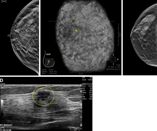
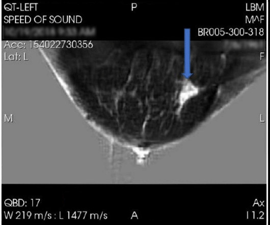
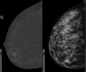
Supplemental breast ultrasound may have utility in imaging women with dense breasts and high risk of advanced or invasive breast cancer, a study published August 6 in Radiology found. Sample images show cancer detection at supplemental ultrasound screening after screening mammography with a negative result. (A)
While services for breast and lung cancer screening were temporarily halted, imagers in x-ray, lung ultrasound, and PET/CT were busy examining patients who presented with COVID-19. The guidelines included best practices for imaging with CT versus chest x-ray in diagnosing COVID, as well as whether imaging is necessary at all in some cases.
Gina Curry (D-Delaware) and would eliminate costs for women for supplemental imaging such as breast MRIs and ultrasounds. When a mammogram reveals an abnormality or an individual is at a higher risk of breast cancer, diagnostic and supplemental imaging is required to determine if the patient needs a biopsy. Komen, in a news release.
While her mammogram yielded negative results, a subsequent ultrasound found breast cancer. Two barriers include access to follow-up imaging such as MRI and ultrasound, and insurance coverage for supplemental imaging. It is so much easier to implement because it can generally be a software upgrade to a mammography machine.
(B) Axial subtracted contrast-enhanced fat-suppressed T1-weighted image from a subsequent-round abbreviated MRI examination performed two years later shows a new 5-mm enhancing mass in the upper outer right breast (arrow), which was not seen on a mammogram performed five months prior. The exam was assessed as BI-RADS category 5. PPV2 21.3%
A team led by Joao Horvat, MD, from the Memorial Sloan Kettering Cancer Center in New York found that CEM depicted 90% of breast cancers compared with 10% on low-energy mammograms alone and 50% on low-energymammogramswith whole-breast ultrasound. RSNA The team identified nine screen-detected cancers diagnosed in eight women.
A team led by Julie Hamzah, MBBS, from Singapore General Hospital, found that symptomatic first breast cancers, dense breasts, and the presence of trabecular thickening on mammography are tied to mammogram detection failure of ipsilateral second breast cancers.
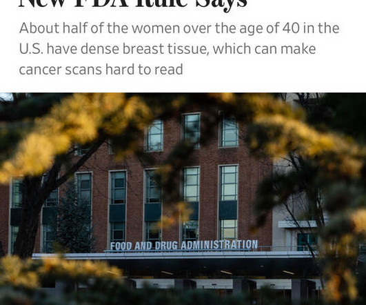
What’s more, the USPSTF concluded that there was insufficient evidence to recommend supplemental screening with MRI or ultrasound in women, regardless of breast density. Food and Drug Administration (FDA) requirement that all women having mammograms receive notice that their breasts are dense or not dense.
House Bill 2411 was introduced in the state by Representative David Cook (R-Globe) and includes eliminating costs for patients for MRI, ultrasound, and diagnostic mammograms. However, out-of-pocket costs for patients can range from $234 for a diagnostic mammogram to more than $1,000 for a breast MRI, according to the organization.
While such diagnosis technology can be used to help doctors diagnose breast cancer, the researchers also emphasized that “there is still much to be improved.” Sereshkeh's group explored the potential of a CNN-based deep learning model that incorporates deep features resulting from mammography staging. International.
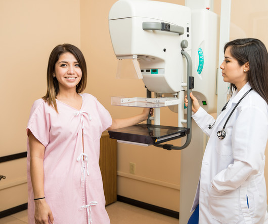
The American Cancer Society recommends starting annual mammogram screenings at age 40. The list below are imaging exams used to diagnose breast cancer. Mammogram Screening Mammogram: Screening mammograms take 2 or more images of each breast. The list below are imaging exams used to diagnose breast cancer.
The earlier breast cancer is diagnosed, the easier it is to treat. Screenings for Breast Cancer The American Cancer Society estimates that more than 252,000 new cases of invasive breast cancer will be diagnosed in women in the U.S. To that end, you should be aware of the common signs and symptoms of breast cancer. this year alone.
While fewer than 5% of women with breast cancer are diagnosed before the age of 40(1), those cancers are usually aggressive, and the young patients suffer from poor survival outcomes. Unfortunately, routine screening mammograms are not recommended for women under 40 because risks outweigh potential benefits at this young age.
At PURE Mammography, we offer 3D breast mammography as well as ultrasound imaging for breasts and other areas of the body. In addition to mammograms and ultrasounds, women should know about the various other breast screenings that are performed. Breast Ultrasound A breast ultrasound is different than an x-ray.
It might be an MRI, an ultrasound, a mammogram, or another imaging test. When a primary care doctor orders specialized testing, say for a patient who complains of breast pain, they may not know the best imaging test to choose.
The product can be used to visualize known or suspected lesions of the breast in adults, as an adjunct to mammography and/or ultrasound. million women diagnosed with breast cancer globally, according to the World Health Organization (WHO).4 Breast Density on a Mammogram. women older than 40 with dense breasts.2
Every woman in the United States who has a mammogram will be notified of her breast density, enabling her to be proactive in obtaining additional imaging to evaluate her breast tissue.
With an X-ray, Ultrasound, Mammogram and CT scan at our disposal, we needed to have a radiologist who could read all these modalities, and give us results in the shortest time possible to enable us to give the best medical care possible” she says. MHRG devised a 24/7 remote radiology solution for the Clinic.
Women with dense breasts are BOTH more likely to develop breast cancer and more likely to have that cancer missed on a mammogram [5] Fig. 1 – Cancer on a mammogram of a fatty vs a dense breast What is Dense Breast Tissue? Breast density is determined through a mammogram and described as one of four categories (Fig.
A trained onsite sonologist with remote interpretation of the ultrasound studies helped them to fulfil their vision. Lives MHRG have touched: “A self-referred 51-year-old with a cough who requested a chest x-ray and a mammogram to be diagnosed with metastatic breast cancer having been treated for pneumonia over and over again…. “45-year-old
Hence, mammograms carried out anywhere can now be viewed by expert breast radiologists in any part of the world thanks to teleradiology. If the breast is dense on the mammogram, an ultrasound must also be carried out. most breast cancers diagnosed after age 50. Age 40-50 Once in 2 years 3. Getting older.-most
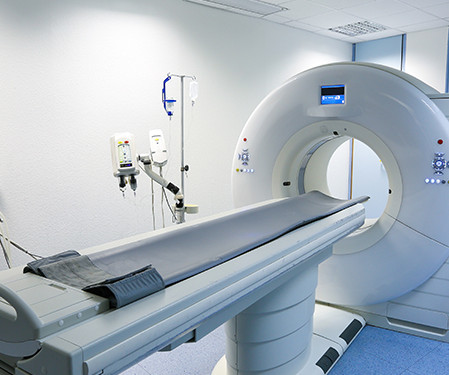
Doctors use imaging tests to see inside a patient’s body and diagnose their illnesses and injuries. There are several types of imaging tests that physicians use to detect cancer in patients: X-Ray, Computed Tomography (CT), Magnetic Resonance Imaging (MRI), Ultrasound (US), Nuclear Medicine, and Positron Emission Tomography (PET).
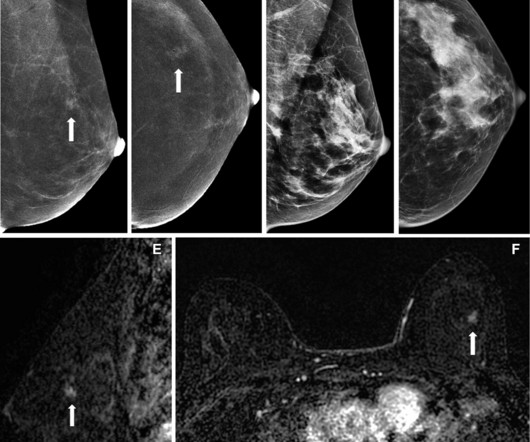
A) Mammogram MLO view. Mammogram CC view. A mammogram demonstrated focal asymmetries involving most of the anterior and mid right breast with diffuse skin thickening, trabecular coarsening, increased overall density, and enlarged right axillary lymph nodes. What is the diagnosis? Xray of the Week Figure 1.
All of the annual scheduled services such as mammograms can now be scheduled, as well as imaging prescribed by physicians for the care of their patients. Patients may also schedule mammograms directly at our facilities in these same locations. Christopher Newman, Chief Medical Officer of Mary Washington Healthcare.
When it comes to accurate diagnoses and effective patient care, getting a second opinion on imaging results can make all the difference. Our abdominal imaging specialists ensure accurate diagnoses for conditions like pancreatic cancer or complex GI issues.
Women diagnosed with fibrocystic breasts may be treated with hormonal birth control or other hormone therapy. PURE Mammography is committed to providing the most efficient mammograms and breast ultrasounds in the most relaxing environment possible. In some cases, if a cyst is very painful, the doctor may drain it.
The theoretical basis for ultrasound physics has been around since 1794, but it wasn’t until 1942, when Dr Karl Theodore Dussik in Austria transmitted an ultrasound beam through a human skull to view the brain, that ultrasound was first used in medicine. (13) This was a defining publication in the field of medical ultrasound. (14)
Breast cancer is the most commonly diagnosed cancer among women, with one in eight facing diagnosis in their lifetime.1 1 Regular mammograms are critical, but their effectiveness is reduced in women with dense breast tissue, who are four to six times more likely to develop breast cancer.2
Breast cancer is currently the most commonly diagnosed cancer among American women – with about one in eight facing a diagnosis in their lifetime. [1] For some patients, tissues can overlap potentially hiding signs of breast cancer; in other cases, the tissue and cancer can show up as white on a mammogram making diagnosis more difficult.
The USPSTF also said that there was insufficient evidence to recommend supplemental screening with MRI or ultrasound in women, regardless of breast density. Gordon added that the guidelines could make way for more interval cancers diagnosed at advanced stages in younger women.
Breast cancer remains the most commonly diagnosed cancer among women in the U.S. i Thankfully, the field of breast screening is not static, and advancements are revolutionizing how we detect and diagnose the disease. Ultrasound rounds out the radiologist’s toolkit for supplemental imaging of women with dense breasts.
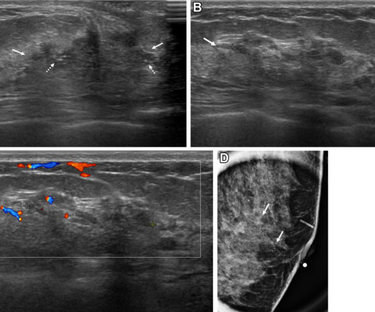
Some nonmass breast lesion characteristics on ultrasound exams can be considered suspicious for malignancy at screening ultrasound, according to research published November 5 in Radiology. Images depict a 43-year-old woman with a malignant non-mass lesion that was diagnosed as microinvasive ductal carcinoma. (A) cm vs. 1.9
We organize all of the trending information in your field so you don't have to. Join 5,000 users and stay up to date on the latest articles your peers are reading.
You know about us, now we want to get to know you!
Let's personalize your content
Let's get even more personalized
We recognize your account from another site in our network, please click 'Send Email' below to continue with verifying your account and setting a password.


Let's personalize your content