This site uses cookies to improve your experience. To help us insure we adhere to various privacy regulations, please select your country/region of residence. If you do not select a country, we will assume you are from the United States. Select your Cookie Settings or view our Privacy Policy and Terms of Use.
Cookie Settings
Cookies and similar technologies are used on this website for proper function of the website, for tracking performance analytics and for marketing purposes. We and some of our third-party providers may use cookie data for various purposes. Please review the cookie settings below and choose your preference.
Used for the proper function of the website
Used for monitoring website traffic and interactions
Cookie Settings
Cookies and similar technologies are used on this website for proper function of the website, for tracking performance analytics and for marketing purposes. We and some of our third-party providers may use cookie data for various purposes. Please review the cookie settings below and choose your preference.
Strictly Necessary: Used for the proper function of the website
Performance/Analytics: Used for monitoring website traffic and interactions
While services for breast and lung cancer screening were temporarily halted, imagers in x-ray, lung ultrasound, and PET/CT were busy examining patients who presented with COVID-19. Her team employed a tracking mechanism for patients who were due for their mammograms once screening operations resumed.
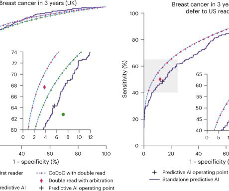
A team of AI and medical specialists working with or for Google Research and Google DeepMind, has developed an AI based system designed to judge the confidence level of existing AI systems used for analyzing medical scans as a means of improving analysis of diagnostic tools, such as mammograms or chest X-rays.
The American Cancer Society recommends starting annual mammogram screenings at age 40. The list below are imaging exams used to diagnose breast cancer. Mammogram Screening Mammogram: Screening mammograms take 2 or more images of each breast. The list below are imaging exams used to diagnose breast cancer.
It is estimated that 1 out of every 8 women in the United States will be diagnosed with breast cancer in her lifetime. One question our technologists are asked frequently is, “What’s the difference between a diagnostic mammogram and a screening mammogram?” first appeared on Clermont Radiology.
Teleradiology-in-Flat-World Introduction : In the realm of modern medicine, diagnostic tools such as X-Rays, mammograms, and CAT scans play a crucial role in identifying and understanding various health conditions.
Adjustments to the Medicare Physician Fee Schedule have historically impacted imaging services, including reductions in the professional component payments for certain procedures like X-rays and MRIs. Improving Patient Outcomes: Accurate and timely diagnoses lead to better treatment plans and improved patient care.
In addition to mammograms and ultrasounds, women should know about the various other breast screenings that are performed. It is an x-ray that takes two or more images of each breast to observe the quality of tissue. Diagnostic mammography may be performed on patients whose routine mammogram detected abnormalities.
The fact is, 75% of women diagnosed have no family history of breast cancer.If your family history is free of breast cancer, you should not neglect a yearly mammogram. Bottom line, if you are a woman over the age of 40, you should be getting a yearly mammogram. Myth: Mammograms can cause cancer.
Through the use of iodine-based x-ray contrast agents, CEM can allow for better visualization of abnormalities in breast tissue that may not be visible with standard mammography.3 million women diagnosed with breast cancer globally, according to the World Health Organization (WHO).4 Breast Density on a Mammogram.
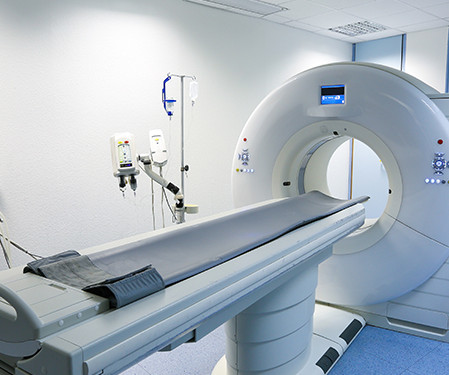
Doctors use imaging tests to see inside a patient’s body and diagnose their illnesses and injuries. There are several types of imaging tests that physicians use to detect cancer in patients: X-Ray, Computed Tomography (CT), Magnetic Resonance Imaging (MRI), Ultrasound (US), Nuclear Medicine, and Positron Emission Tomography (PET).
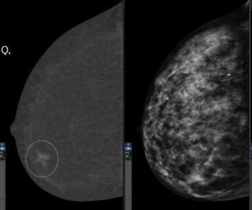
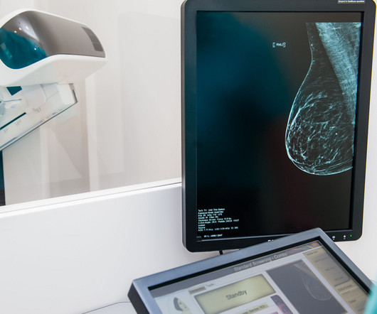
Breast cancer is currently the most commonly diagnosed cancer among American women – with about one in eight facing a diagnosis in their lifetime. [1] For some patients, tissues can overlap potentially hiding signs of breast cancer; in other cases, the tissue and cancer can show up as white on a mammogram making diagnosis more difficult.
With an X-ray, Ultrasound, Mammogram and CT scan at our disposal, we needed to have a radiologist who could read all these modalities, and give us results in the shortest time possible to enable us to give the best medical care possible” she says. She has since received definitive treatment and is doing well.
All of the annual scheduled services such as mammograms can now be scheduled, as well as imaging prescribed by physicians for the care of their patients. Patients may also schedule mammograms directly at our facilities in these same locations. Christopher Newman, Chief Medical Officer of Mary Washington Healthcare.
Lives MHRG have touched: “A self-referred 51-year-old with a cough who requested a chest x-ray and a mammogram to be diagnosed with metastatic breast cancer having been treated for pneumonia over and over again…. “45-year-old
Mammography is an X-Ray of the breasts carried out on a machine dedicated to it because the breast is not of uniform thickness and requires a dedicated unit. Hence, mammograms carried out anywhere can now be viewed by expert breast radiologists in any part of the world thanks to teleradiology. Age 40-50 Once in 2 years 3.
Whether it’s X-rays, MRIs, mammograms , CT scans, or other subspecialty , our streamlined process ensures swift delivery of results without compromising on quality. Sources: Ncbi.nlm.nih.gov ama-assn.org Openai.com The post Healthcare Services for Native Americans (IHS) first appeared on Vesta Teleradiology.
Women with dense breasts are BOTH more likely to develop breast cancer and more likely to have that cancer missed on a mammogram [5] Fig. 1 – Cancer on a mammogram of a fatty vs a dense breast What is Dense Breast Tissue? Breast density is determined through a mammogram and described as one of four categories (Fig.
When it comes to accurate diagnoses and effective patient care, getting a second opinion on imaging results can make all the difference. Our abdominal imaging specialists ensure accurate diagnoses for conditions like pancreatic cancer or complex GI issues. This builds trust with referring physicians and patients.
Tucked away beneath all of the symbolism and public events, is the quiet experience of the mammogram and the radiology technology that makes it possible. Why are mammograms at the center of this public health battle? Why is a Mammogram Important? Here’s what you need to know. The short answer is detection.
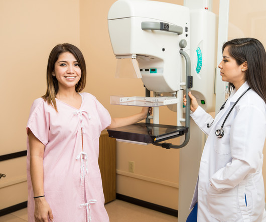
Screening Mammograms have proven to be essential for the early detection of breast cancer. What Is A Screening Mammogram? A mammogram is an examination that uses a special low dose X-Ray machine to evaluate breast tissue. The mammogram unit is designed specifically for breast imaging.
A combination of canceled elective screenings and procedures, staff and PPE shortages, office closures, and personal health concerns all contributed to a decline in the number of routine mammograms provided for at-risk women. The result is a rise in breast cancer diagnoses. Why do I need a Mammogram Every Year?
That is, it is diagnosed after a test whose result was negative and before the next evaluation. Conversely, if the number of tumors diagnosed after screening is low, it indicates that the program is fulfilling its function: to detect this disease at an early stage. per thousand people, while in the second it was 0.93.
You’re more likely to be diagnosed with breast cancer than any other cancer (besides skin cancer). That decline has been attributed, in large part, to annual screening mammograms. Each year, during your mammogram appointment, a full assessment is done to determine if you have one or multiple risk factors.
It all started when Wilhelm Conrad Röntgen discovered X-rays in 1895. After working for weeks in his lab experimenting on the production of ‘strange rays’, which he referred to as ‘X’, he asked his wife Anna Bertha to lend ‘a hand’, the left one to be precise, which he used to produce the first X-ray image.
Recently, the use of breast thermography as a screening tool for breast cancer has come into the picture as an alternative to the standard screening mammogram. Mammography is a low dose x-ray used to image the soft tissue of the breast. What is Thermography? What is Mammography? Mammography uses a low dose of radiation.
Mammography Mammography is a specialized imaging service that uses low-dose X-rays to examine breast tissue. Regular mammograms can detect tumors that are too small to be felt, significantly increasing the chances of successful treatment. Non-Invasive: The procedure is safe, quick, and involves minimal radiation exposure.
We organize all of the trending information in your field so you don't have to. Join 5,000 users and stay up to date on the latest articles your peers are reading.
You know about us, now we want to get to know you!
Let's personalize your content
Let's get even more personalized
We recognize your account from another site in our network, please click 'Send Email' below to continue with verifying your account and setting a password.


Let's personalize your content