This site uses cookies to improve your experience. To help us insure we adhere to various privacy regulations, please select your country/region of residence. If you do not select a country, we will assume you are from the United States. Select your Cookie Settings or view our Privacy Policy and Terms of Use.
Cookie Settings
Cookies and similar technologies are used on this website for proper function of the website, for tracking performance analytics and for marketing purposes. We and some of our third-party providers may use cookie data for various purposes. Please review the cookie settings below and choose your preference.
Used for the proper function of the website
Used for monitoring website traffic and interactions
Cookie Settings
Cookies and similar technologies are used on this website for proper function of the website, for tracking performance analytics and for marketing purposes. We and some of our third-party providers may use cookie data for various purposes. Please review the cookie settings below and choose your preference.
Strictly Necessary: Used for the proper function of the website
Performance/Analytics: Used for monitoring website traffic and interactions
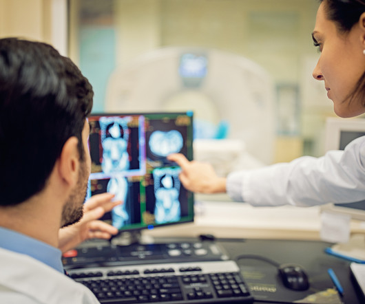
Multiparametric MRI (mpMRI) is a widely used approach for diagnosing patients with prostate cancer and reduces the need for invasive biopsies in approximately 30% of cases, Brembilla explained. Our preliminary data confirmed the potential increase of sensitivity of the combined use of PSMA PET with MRI compared to each modality alone.
F-18 FAPI-PET/CT is superior to F-18 FDG-PET/CT for diagnosing and staging patients with pancreatic cancer, according to a study published January 4 in the Journal of NuclearMedicine. Image courtesy of the Journal of NuclearMedicine. Tumor is marked by arrows. DWI = diffusion-weighted imaging.
PET/MRI imaging shows promise in diagnosing fevers or inflammation of unknown origin and may have advantages over PET/CT, according to a study published January 3 in the European Journal of Radiology. This imaging technique has especially high sensitivity, negative predictive value and low radiation exposure,” the group concluded.
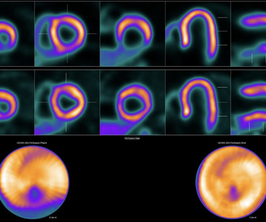
Di Carli noted that currently, SPECT MPI – also a nuclearmedicine imaging exam, but one that requires higher radiation doses to patients and produces less resolution – remains the workhorse in the field. The game-changer could be the pending U.S. approval of new PET radiotracers, such as F-18 flurpiridaz , he said.
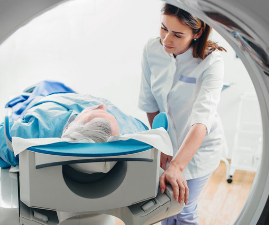
Nuclearmedicine is a form of specialty medicine that uses radioactive tracers to evaluate bodily functions and to diagnose and treat a wide range of health conditions. Nuclear scans produce images of the body’s anatomy that cannot be obtained as clearly or fully with other imaging techniques.
The World Health Organization (WHO) on September 17 will offer a webinar on the importance of accurate and timely diagnoses in ensuring patient safety that will include discussion of medical imaging and radiation exposure. to 1:30 p.m. to 1:30 p.m. to 1:30 p.m. Central European Time.
Morphological markers on MRI help diagnose autism. Effects of low-dose ionizing radiation on genomic instability in interventional radiology workers. Luthria, et al, Journal of NuclearMedicine , October 26, 2023. Rankine, et al, International Journal of Radiation Oncology, Biology, Physics , September 1, 2023.
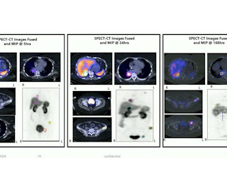
PET/CT imaging with a new gallium-68-based prostate cancer radiotracer shows promise for detecting recurrent metastatic disease, according to a study published February 20 in the Journal of NuclearMedicine. All patients had undergone either radical prostatectomy or radiation therapy. (A)
R)(N)(ARRT) About the Honorees Arianna Apodaca is a radiation therapist at Austin Cyberknife , a cancer treatment center in Austin, Texas. At age 19, Ariana was diagnosed with Stage II Triple Negative Breast Cancer. Patrick’s efforts have led to an impressive level of quality and consistency in nuclear imaging across the enterprise.
His accidental discovery forever changed medicine and it has been a key tool in diagnosing and treating injuries and diseases for 128 years.” ASRT also noted it represents 157,000 members who perform medical imaging procedures or plan and deliver radiation therapy, and is the largest radiologic science association in the world.
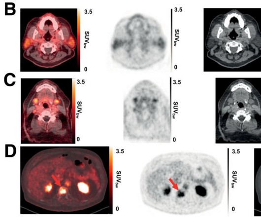
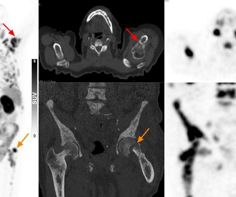
milla1cf Tue, 08/15/2023 - 16:28 August 15, 2023 — In elderly patients with suspected prostate cancer, PSMA PET/CT can diagnose advanced disease and aid in therapy selection without the need for a biopsy.
The college represents thousands of radiologists, radiation oncologists, nuclearmedicine physicians, and physicists. This useful medical imaging helps physicians diagnose or assess any number of conditions, injuries, or pathologies. Today, the College has 54 chapters.
We can’t treat what we don’t see, which is why we require precise image quality to help diagnose, plan treatment for, and monitor disease,” explains Prof. Flavio Forrer , MD, PhD, Chairman of NuclearMedicine in the Division of Radiology and NuclearMedicine at Kantonsspital St. Gallen in Switzerland [vii].
professor of the Department of Radiation Oncology and the Department of Immunology and Theranostics at City of Hope , and principal investigator of the RefleXion-supported PyL imaging study. professor and chair of City of Hope’s Department of Radiation Oncology. “We
The collaboration seeks to better equip clinicians with technology to precisely diagnose and treat medical conditions and personalize the patient experience at each step of their care journey through the clinical translation of novel technologies and approaches to medical imaging and theranostics.
GE HealthCare expects to integrate MIM Software solutions into its advanced visualization offerings to facilitate AI-based segmentation and contouring as well as dosimetry analysis for patients across their treatment journeys and in the growing fields of radiology, molecular imaging, and radiation oncology.
Although the word “radiology” sounds like it involves radiation, that is not always the case – for example, MRI (magnetic resonance imaging) and ultrasound do not use radiation in their medical imaging technologies. Digital X-rays use less radiation and are employed for the same purposes.
In 2011, a large study examined the use of x-rays and other radiation imaging on children—they estimated that the average child would get more than seven radiation scans by the age of 18. No doubt, then, that the role of a pediatric radiologist is important in accurately diagnosing and treating diseases and conditions in children.
MRI-Scan-Teleradiology Description: Radiology is a crucial medical specialty that plays a fundamental role in diagnosing and monitoring a wide range of medical conditions. Introduction to Radiology : Radiology is a branch of medicine that uses medical imaging techniques to diagnose and treat diseases and injuries.
This is just one variation between adult and pediatric anatomy, but there are hundreds more that can make a big difference in radiologic diagnoses. One additional consideration when imaging pediatric patients is their exposure to ionizing radiation. In this instance, the Radiologist terminated the exam after this single view.
Imaging services are an essential way to accurately diagnose injuries, medical conditions, and diseases. High-definition images provide precise information that allow physicians to accurately diagnose a variety of conditions. What Is An Outpatient Imaging Center? This helps doctors recommend appropriate and effective treatments.
Theranostics pairs diagnostic biomarkers that can be visualized on nuclearmedicine imaging with therapeutic agents that share a specific target in diseased cells or tissues. After binding to the receptor, the drug works by entering the cell allowing radiation to cause damage to the tumor cells.
It is one of the first scans performed on patients, and the information is used to diagnose and evaluate cancer-related complications, including malignancy, obstruction, and infection. Radiomics provides a wealth of information, pulling from CTs, MRIs, and PETs, connecting imaging with precision medicine.
The model performed lowest on image-containing questions in the nuclearmedicine domain, correctly answering only 2 of 10 questions. We noted an alarming tendency for the model to provide correct diagnoses based on incorrect image interpretations, which could have significant clinical implications.”
Theranostics pairs diagnostic biomarkers that can be visualized on nuclearmedicine imaging with therapeutic agents that share a specific target in diseased cells or tissues. After binding to the receptor, the drug works by entering the cell allowing radiation to cause damage to the tumor cells.
Several presenters at the Society for NuclearMedicine and Molecular Imaging's (SNMMI) 2024 meeting in Toronto shared new findings in women's studies. and included limited single and cumulative radiation doses. In the newly diagnosed cohort 1, standard of care imaging detected 12/14; FES detected 11/14 (P > 0.99).
18F-flurpiridaz PET MPI obtained images at a lower radiation dose than 99Tc-SPECT MPI and performed similarly in both obese and non-obese patients. “Due This can result in inferior image quality and diagnostic performance despite requiring a higher dose of radiation.” Of the 578 patients with evaluable studies, 298 (51.6
Patients may bring legal charges against radiologists or their imaging facility for a number of reasons including failure to diagnose breast cancer or negligence due to a fall resulting in injury. Administrators and Support Staff: receptionists, nurses, medical physicist, radiation officer, etc. provide the logistical backbone.
We organize all of the trending information in your field so you don't have to. Join 5,000 users and stay up to date on the latest articles your peers are reading.
You know about us, now we want to get to know you!
Let's personalize your content
Let's get even more personalized
We recognize your account from another site in our network, please click 'Send Email' below to continue with verifying your account and setting a password.


Let's personalize your content