This site uses cookies to improve your experience. To help us insure we adhere to various privacy regulations, please select your country/region of residence. If you do not select a country, we will assume you are from the United States. Select your Cookie Settings or view our Privacy Policy and Terms of Use.
Cookie Settings
Cookies and similar technologies are used on this website for proper function of the website, for tracking performance analytics and for marketing purposes. We and some of our third-party providers may use cookie data for various purposes. Please review the cookie settings below and choose your preference.
Used for the proper function of the website
Used for monitoring website traffic and interactions
Cookie Settings
Cookies and similar technologies are used on this website for proper function of the website, for tracking performance analytics and for marketing purposes. We and some of our third-party providers may use cookie data for various purposes. Please review the cookie settings below and choose your preference.
Strictly Necessary: Used for the proper function of the website
Performance/Analytics: Used for monitoring website traffic and interactions
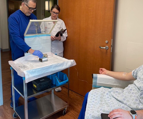
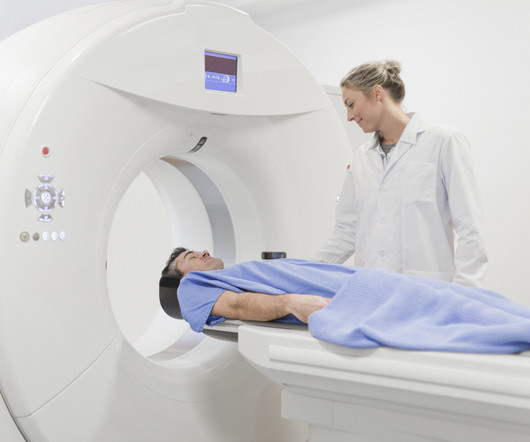
The L block lead shield over the syringe helps protect certified nuclear medicine technologist James Johnson (left), and nuclear radiologist Penny Vroman, MD (right), from radiation emitted by the drug. After binding to the receptor, the drug works by entering the cell allowing radiation to cause damage to the tumor cells.
Teleradiology & Radiology data for artificial intelligence (AI) Introduction: Embark on a journey into the world of medical imaging as we unravel the distinctions between two powerful diagnostic tools—Computed Tomography (CT) scans and Positron Emission Tomography (PET) scans.
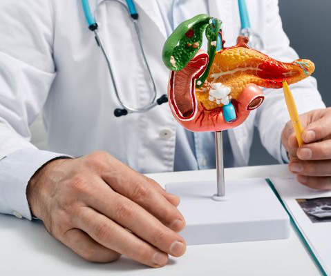
Depending on the disease’s progression, your doctor may recommend surgery, chemotherapy, radiation therapy or a combination of these treatments. This procedure is less invasive than other methods and provides a higher level of detail compared to traditional X-rays, while also reducing radiation exposure.
The L block lead shield over the syringe helps protect certified nuclear medicine technologist James Johnson (left), and nuclear radiologist Penny Vroman, MD (right), from radiation emitted by the drug. After binding to the receptor, the drug works by entering the cell allowing radiation to cause damage to the tumor cells.
We can’t treat what we don’t see, which is why we require precise image quality to help diagnose, plan treatment for, and monitor disease,” explains Prof. Medical imaging is a crucial tool for diagnosing disease, identifying a course of treatment, and determining whether therapy is successful for millions of patients around the world.
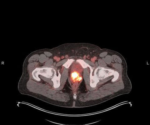
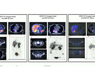
milla1cf Tue, 05/02/2023 - 23:50 May 2, 2023 — Blue Earth Diagnostics , a Bracco company and recognized leader in the development and commercialization of innovative PET radiopharmaceuticals, today announced additional results from its completed Phase 3 SPOTLIGHT trial of 18F-rhPSMA-7.3 18F-rhPSMA-7.3 on behalf of the SPOTLIGHT Study Group.
Results demonstrated high detection rates (% positive PETscans) even at low PSA levels. in Newly Diagnosed Prostate Cancer Blue Earth Diagnostics Announces Results on Clinical Factors Impacting Detection Rates from Phase 3 SPOTLIGHT Trial of Investigational PET Imaging Agent 18F-rhPSMA-7.3
It allows physicians to diagnose and treat many different health conditions, injuries, and diseases more accurately and easily. A CT (computerized tomography) scan is used to enable doctors to see clear images of the structures and tissues of your body. Diagnostic imaging plays a very important role in modern medicine.
tesla MRI AI body composition analysis Cardiac PET Cryo/thermoablation CT colonography Genicular artery embolization Hyperpolarized xenon-129 MRI PET/MRI Photon-counting CT Radiomics Theranostics Whole-body MRI screening Image of the Year 3D PET/MR image. Morphological markers on MRI help diagnose autism.
and included limited single and cumulative radiation doses. The study examined tumor uptake of Ga-68-NeoB on 26 scans in 29 patients: 10 from patients with newly diagnosed breast cancer, 16 from patients with persistent or recurrent disease, and three from patients without clinical verification of an active tumor.
We organize all of the trending information in your field so you don't have to. Join 5,000 users and stay up to date on the latest articles your peers are reading.
You know about us, now we want to get to know you!
Let's personalize your content
Let's get even more personalized
We recognize your account from another site in our network, please click 'Send Email' below to continue with verifying your account and setting a password.


Let's personalize your content