This site uses cookies to improve your experience. To help us insure we adhere to various privacy regulations, please select your country/region of residence. If you do not select a country, we will assume you are from the United States. Select your Cookie Settings or view our Privacy Policy and Terms of Use.
Cookie Settings
Cookies and similar technologies are used on this website for proper function of the website, for tracking performance analytics and for marketing purposes. We and some of our third-party providers may use cookie data for various purposes. Please review the cookie settings below and choose your preference.
Used for the proper function of the website
Used for monitoring website traffic and interactions
Cookie Settings
Cookies and similar technologies are used on this website for proper function of the website, for tracking performance analytics and for marketing purposes. We and some of our third-party providers may use cookie data for various purposes. Please review the cookie settings below and choose your preference.
Strictly Necessary: Used for the proper function of the website
Performance/Analytics: Used for monitoring website traffic and interactions
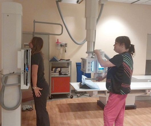
12, 2025 Konica Minolta Healthcare Americas, has published a case study by clinicians in the pulmonary and radiology departments at ASST Fatebenefratelli Sacco (Milan, Italy) demonstrating the use of Dynamic Digital Radiography (DDR) to help definitively diagnose diaphragm dysfunction. tim.hodson Fri, 02/14/2025 - 15:14 Feb.12,
Louis asked, “How much ionizing radiation are neonatal patients exposed to during interventional procedures?” Radiation doses estimated in infants with congenital heart disease Monday, November 27 | 3:10 p.m.-3:20 In one, a group at the University of Washington in St. 3:20 p.m. | 8:30 a.m. | 9:20 a.m. | 1:50 p.m. | 1:50 p.m. |
Chest dynamic digital radiography (DDR) may have received a boost toward clinical use in patients with lung disorders, with researchers developing AI to perform time-consuming analysis involved in the technology, according to researchers in New York City. Ultimately, DDR has unique advantages over traditional studies, the authors noted.
Morphological markers on MRI help diagnose autism. Effects of low-dose ionizing radiation on genomic instability in interventional radiology workers. Quantifying Regional Radiation-Induced Lung Injury in Patients Using Hyperpolarized 129Xe Gas Exchange Magnetic Resonance Imaging. Shah, et al, Radiography , January 9, 2024.
Understanding the distinction between radiography and sonography is vital in the increasingly specialized and multifaceted world of medical imaging. Radiography uses ionizing radiation to capture detailed internal images, which helps diagnose various conditions.
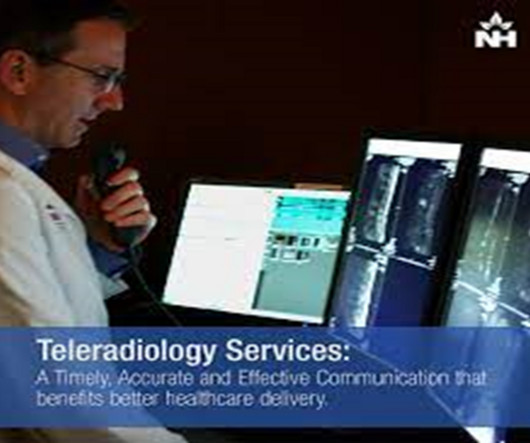
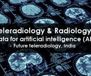
Teleradiology Introduction: Dental radiography plays a pivotal role in modern dentistry, enabling practitioners to diagnose and treat various oral health conditions. However, the field of dental radiography has undergone significant advancements, along with its own set of challenges and triumphs.
MRI-Scan-Teleradiology Introduction: Dental radiography is an essential component of modern dentistry, offering valuable insights into oral health and guiding treatments with precision. Patient-Centered Approach: Dental radiography starts with the patient. This quality control is vital for accurate diagnoses.
X-ray radiography is a noninvasive diagnostic method that uses X-rays—electromagnetic radiation—to produce images of the body's internal structures. Essential in medical fields for diagnosing injuries and diseases and monitoring treatment progress, X-ray radiography is a critical tool in patient care and medical decision-making.
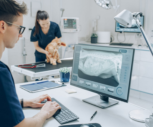
Their expertise in handling animals, positioning them correctly, and ensuring optimal image quality is essential for accurate diagnoses. Poor positioning can lead to retakes, increased radiation exposure, and misdiagnoses. Diagnostic Imaging Systems has made it easy for you to purchase radiation protection online.
Introduction: The world of radiography is one filled with immense responsibility and precision, where the journey to capture images of the human body’s inner workings is not without its challenges. Radiation Safety: A Top Priority: Radiation safety is paramount in radiography.
Chest radiography is a common diagnostic tool, but significant training and experience is required to interpret exams correctly,” said lead researcher Louis L. Too many false-positive diagnoses would result in unnecessary imaging, radiation exposure and increased costs.” Plesner, M.D., resident radiologist and Ph.D.
Radiography soon became a vital tool in medicine, and for decades, it relied on photographic film to capture and develop X-ray images. The Birth of Digital Radiography: The 21st century brought a seismic shift as X-ray imaging transitioned from film to digital radiography.
Chapter 2: The Art and Science of Radiography A closer look at the development of radiography, the first X-ray imaging method. How radiography has played a pivotal role in diagnosing bone fractures, identifying foreign bodies, and shaping early healthcare practices.
From their serendipitous discovery to their pivotal place in diagnosing hidden conditions, we will illuminate the invisible to understand the lasting impact of X-rays. Chapter 3: The Evolution of Radiography: From Shadows to Images An exploration of the development of radiography, the earliest X-ray imaging technique.
From its early discovery to its integral position in diagnosing hidden conditions, we will uncover the layers beneath the surface to appreciate the indispensable role of X-ray imaging in patient care. How radiography transformed healthcare by allowing the visualization of internal structures.
From Film to Digital: The transition from film-based X-ray systems to digital technology marked a turning point in dental radiography. Digital sensors and intraoral cameras offer instant image acquisition, reduced radiation exposure, and the ability to enhance and manipulate images for better diagnostics.
Angela Young explains how the process of making a podcast helped not only others with a diagnosed brain tumour but gave comfort and support to herself as she embarked on a course of radiotherapy. After all, positivity radiates. It can also make you adjust your priorities in life.
Yet a lack of access to high-quality imaging can lead to delayed or missed diagnoses, further exacerbating the health disparities experienced by people who live in disadvantaged or remote communities. Low-dose X-ray Solutions Serve Broadest Patient Population Digital radiography has come a long way at Massac Memorial as well.
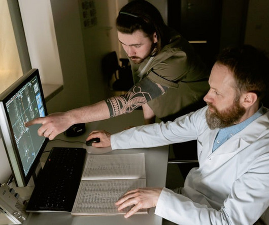
This blog delves into the essential role played by X-ray technicians in the process of visualizing health, capturing images that hold the key to diagnosing and treating medical conditions. It’s the foundation upon which many diagnoses are made, a tool that unveils the hidden aspects of our health.
The historical backdrop of Wilhelm Roentgen’s serendipitous discovery and the birth of radiography. Chapter 3: Types of X-ray Technology: Beyond Radiography An exploration of the various modalities and applications of X-ray technology, from radiography to fluoroscopy and computed tomography (CT).
Common Indications Scaphoid Fracture A feasible alternative to MDCT for the detection of extremity fractures at a reduced radiation dose. 1 Distal Radius Fracture Post-operatively, CBCT can diagnose scaphoid union at an early follow-up and prevents longer immobilization and interruption of activity or work.
This innovative technology has the potential to change the way we diagnose a range of medical conditions,” according to Norbert Pelc, Sc.D., Planning for Lung Disease Scanning Between late 2023 and early 2024, AIxSCAN, Inc. plans to produce over 50 lung disease patients scans in the U.S. AIxSCAN, Inc.
Chapter 3: The Radiologic Toolbox – Types of X-ray Imaging An exploration of the various types of X-ray imaging, including radiography, fluoroscopy, computed tomography (CT), and more. The importance of minimizing radiation exposure while maintaining diagnostic accuracy.
The principles of radiation and how X-rays interact with the human body to create diagnostic images. Chapter 3: Types of X-ray Imaging: Beyond Radiography An exploration of the various types of X-ray imaging, including radiography, fluoroscopy, and computed tomography (CT).
Radiation Safety: Technologists are responsible for operating the X-ray equipment safely, limiting radiation exposure to patients and themselves. They often wear lead aprons and use shielding devices to minimize radiation exposure. This ensures easy access for the dental team when making diagnoses and treatment decisions.
Film-Based Radiography: Discuss the era of film-based radiography, highlighting its advantages and limitations. The Dawn of Digital Imaging: Explore the transition from film to digital radiology, emphasizing the benefits of digital technology, including immediate results and reduced radiation exposure.
With the advent of cutting-edge technology, digital dentistry is reshaping the way we diagnose, treat, and prevent oral health issues. Digital Radiography: Traditional film-based X-ray imaging is making way for digital radiography.
Insights into their daily work, challenges, and the fulfillment of shaping diagnoses. Chapter 4: Revealing the Unseen – The Power of Imaging in Diagnoses Real-life case studies that illustrate the transformative impact of medical imaging on patient diagnoses.
As radiology departments proliferated in hospitals, X-rays became indispensable for diagnosing a wide range of conditions. Traditional film-based X-rays gave way to digital radiography (DR) and computed radiography (CR). Advanced Imaging Modalities: X-ray technology has expanded beyond conventional radiography.
Trauma : In these injuries one would normally go for a standard radiography followed by second-line imaging such as CT. The possibility of inaccurate or missed diagnoses ranging on average from 20 to 40% in the literature is impossible to ignore and warrants 3D imaging as a primary modality.
These numbers tell us how much radiation was used to create that image and they provide valuable feedback for determining if an image is diagnostic. If the DI is way off, it could mean youve either under or overexposed your image, potentially compromising the diagnostic quality or unnecessarily increasing radiation dose.
We organize all of the trending information in your field so you don't have to. Join 5,000 users and stay up to date on the latest articles your peers are reading.
You know about us, now we want to get to know you!
Let's personalize your content
Let's get even more personalized
We recognize your account from another site in our network, please click 'Send Email' below to continue with verifying your account and setting a password.


Let's personalize your content