This site uses cookies to improve your experience. To help us insure we adhere to various privacy regulations, please select your country/region of residence. If you do not select a country, we will assume you are from the United States. Select your Cookie Settings or view our Privacy Policy and Terms of Use.
Cookie Settings
Cookies and similar technologies are used on this website for proper function of the website, for tracking performance analytics and for marketing purposes. We and some of our third-party providers may use cookie data for various purposes. Please review the cookie settings below and choose your preference.
Used for the proper function of the website
Used for monitoring website traffic and interactions
Cookie Settings
Cookies and similar technologies are used on this website for proper function of the website, for tracking performance analytics and for marketing purposes. We and some of our third-party providers may use cookie data for various purposes. Please review the cookie settings below and choose your preference.
Strictly Necessary: Used for the proper function of the website
Performance/Analytics: Used for monitoring website traffic and interactions
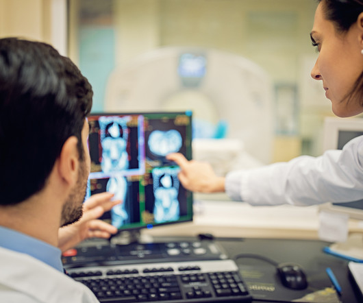
12, 2025 Konica Minolta Healthcare Americas, has published a case study by clinicians in the pulmonary and radiology departments at ASST Fatebenefratelli Sacco (Milan, Italy) demonstrating the use of Dynamic Digital Radiography (DDR) to help definitively diagnose diaphragm dysfunction. tim.hodson Fri, 02/14/2025 - 15:14 Feb.12,
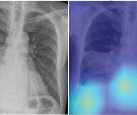
In this AJR accepted manuscript , a DL model was developed in 7,105 patients via one institution from March 2013 to December 2019 (3:1:1 allocation to training, validation, and internal test sets) to predict risk of all-cause mortality within 30 days after CAP diagnosis using patients’ initial chest radiograph. CURB-65 score). Hwang et al.
ChatGPT-4 outperformed human clinicians in determining pretest and post-test disease probability after a negative test result involving chest radiographs and mammograms, according to a research letter published December 11 in JAMA Network Open.
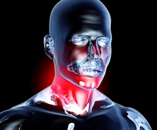
In an analysis of cases where F-18 FDG radiotracer uptake was reported as suspicious for oropharynx cancer on PET/CT scans, the number of patients who were actually diagnosed was low, the group found. Further analysis revealed that these diagnosed patients had higher F-18 FDG mean ipsilateral radiotracer uptake.
a) Raw example of a dynamic digital radiograph. (b) Moreover, while PFTs are vital for diagnosing and monitoring pulmonary disorders, they pose challenges in accessibility, such as in patients with neuromuscular conditions or those experiencing flares of chronic obstructive pulmonary disease, they suggested.
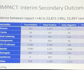
This competition demonstrated the value of AI in detecting and localizing many pathologies in chest radiographs by simulating the real work situations of radiologists,” the group wrote. The study was published February 8 in the Journal of Imaging Informatics in Medicine.
Dubai-based Zscale Labs is launching Neuromorphic AI, an AI software application for multilabel classification of chest radiographs. Neuromorphic AI leverages the firm's hyperdimensional computing and deep-learning technologies to assist radiologists and healthcare providers in diagnosing multiple chest conditions from x-ray images.
Importantly, we are gaining deeper insights into disease processes themselves, which enhances our ability to diagnose and potentially treat conditions that were previously beyond our reach." I'm a radiographer,' " Stewart recalled. I thought, 'I'm not a teacher. But Hennessy encouraged her.
Within the interim results, Woznitza reflected the results as they relate to four categories: time to lung cancer diagnosis, time to CT, time to urgent referral, and time to treatment start in relation to 191 lung cancer cases diagnosed. UCLH has produced 9,217 chest x-rays from 8,072 patients. The other 102 were prioritized.
Teleradiology-India Introduction: “X-ray Visionaries” takes you on a compelling journey to unveil the expertise of radiographers and technologists, the unsung heroes of X-ray technology. Chapter 1: Introduction to Radiographers and Technologists An overview of the pivotal roles radiographers and technologists play in healthcare.
It uses AI to analyze CT scans, X-rays and pathology slides, supporting clinicians in detecting and diagnosing medical conditions faster and more accurately. Averaged across all findings on chest radiographs.) [2]AIDE aims to address the global shortage of 1.5 Lancet Digital Health. Published 2021. 2]AIDE study, Alfred Health.
At seven days post-imaging, nearly half had unreported brain and chest CT scans, while 59% had unreported chest radiographs. For patients, these delays mean longer wait times for diagnoses and treatments. Even at six months, about 20% of these studies remained unreported. The consequences are significant.
Key Points: Weight Bearing (WB) radiographs are limited by 1) their 2D nature and 2) knee positioning. Optimal knee positioning and X-Ray beam angle differs by person and can be unreliable with WB radiographs. Optimal knee positioning and X-Ray beam angle differs by person and can be unreliable with WB radiographs.
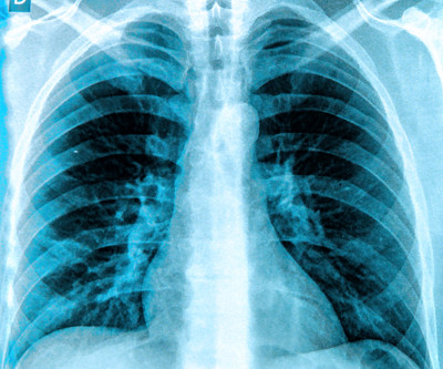
Morphological markers on MRI help diagnose autism. Commercially Available Chest Radiograph AI Tools for Detecting Airspace Disease, Pneumothorax, and Pleural Effusion. Generative Artificial Intelligence for Chest Radiograph Interpretation in the Emergency Department. Cognitive Motor Dissociation in Disorders of Consciousness.
"We identified an association between baseline MRI-defined knee osteoarthritis and the development of incident radiographic and symptomatic disease during up to 11 years of follow-up," the group noted. However, a substantial proportion of individuals with baseline MRI-defined knee OA did not develop radiographic knee OA during follow-up."
The World Health Organization (WHO) on September 17 will offer a webinar on the importance of accurate and timely diagnoses in ensuring patient safety that will include discussion of medical imaging and radiation exposure. to 1:30 p.m. to 1:30 p.m.
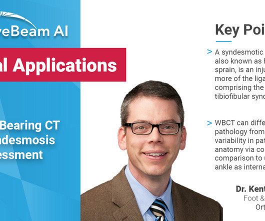
Key Points: Currently plain radiographs are the standard method in diagnosing syndesmotic ankle injuries even though the distal tibiofibular joint cannot be assessed due to superposition of the osseous structures in the foot.
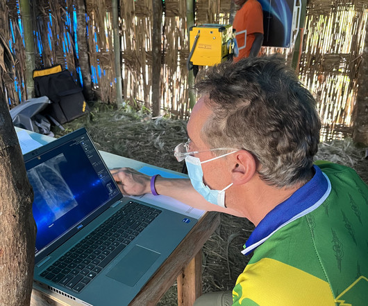
They were able to successfully operate MinXray’s Impact Wireless – a portable and battery-powered digital radiography system – taking several sets of diagnostic radiographic images at the Mount Everest Base Camp in Khumjung, Nepal, at an altitude of 17,598 feet.
Rob Liddell, MD, is a diagnostic radiologist who used MinXray’s Impact Wireless system to take radiographic images of patients in the village and screen for common diseases in the region, such as tuberculosis and emphysema. He also used the system to diagnose cancers, infections and various musculoskeletal injuries.
Non-WB radiographs could not determine instability or severity of midfoot collapse; surgical intervention postponed. One year later, deformity had significantly progressed, but the remaining bone stock was unclear from plain radiographs. The patient was diagnosed with Charcot arthropathy. History of dorsal foot ulcers.
However, the ASRT, like others, has emphasized that RAs (unlike nurse practitioners or physician assistants) are restricted from making diagnoses and must work under the direct supervision of a radiologist.
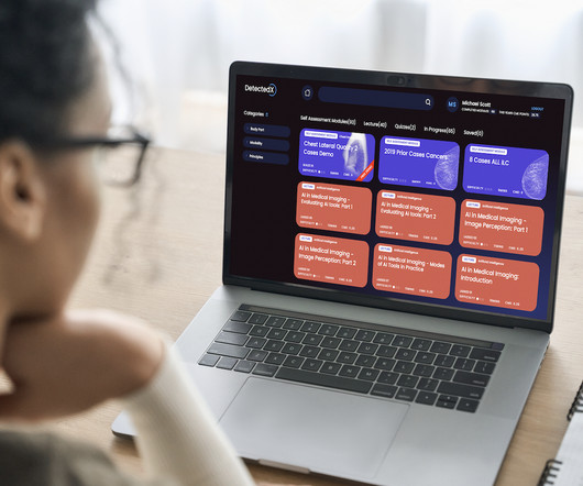
DetectedX was founded to help radiologists and doctors worldwide to diagnose cases of breast cancer, lung cancer, and COVID-19 faster and more accurately. MyImageDx facilitates the delivery of learning world-wide for peer review, MDT and discrepancy activities for students, radiologists and radiographers anywhere, anytime.
Introduction: Delve into the illuminating world of X-rays and their pivotal role in diagnosing and monitoring Inflammatory Bowel Disease (IBD). Diagnosing Inflammatory Bowel Disease with X-Ray: Illuminating the Diagnostic Process Explore how X-rays contribute to the diagnosis of Inflammatory Bowel Disease.
A team led by Avneesh Chhabra, MD, of the University of Texas Southwestern Medical Center in Dallas, found that the system's overall accuracy was higher than final diagnoses rendered without using it, at 65% compared with 55%. The study results were published August 27 in Radiology.
In the third blog of her series on AI and the radiographer, Shamie Kumar explores the impact on the radiographer when AI is integrated within an imaging modality. The question to explore in this blog is when AI is integrated within an imaging modality itself and how that may impact a radiographer.
Patients with ARIA sometimes have headaches, but they are usually asymptomatic and only diagnosable with MRI. “It Most patients with asymptomatic ARIA meeting specific radiographic and clinical criteria may continue to receive treatment. It is essential for the radiologist to recognize and monitor ARIA,” Dr. Agarwal said. “As
Up to 40% of missed diagnoses of ankle trauma associated fractures could theoretically be avoided with primary use of WBCT in accident and emergency departments. Researchers analyzed the limited literature available on using CBCT as a primary modality for diagnosing AS and CLAI.
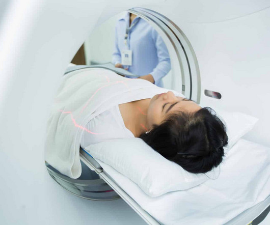
These enable a wide range of diagnostics for conditions that can often not be diagnosed using traditional radiographic imaging techniques. MRIs and CT scans are advanced medical imaging techniques that create detailed images of the internal structures of the body.
Foot X-rays are the most common method podiatrists use to diagnose injuries and other conditions. If a patient is unable to take four steps on their foot without support, that’s a sure sign an X-ray, also called a radiograph, is needed.
Diagnostic imaging tests are tools used by physicians to diagnose a range of medical conditions. Why Diagnostic Imaging Methods Are Important Medical imaging is used to diagnose, monitor, and treat medical problems. X-ray Also called a radiograph, an X-ray uses radiation to create images of the body.
million women diagnosed with breast cancer globally, according to the World Health Organization (WHO).4 RadioGraphics 2019 39:7, 1907-1920. RadioGraphics 2021 41:3, 829-839. 3 About breast cancer and contrast-enhanced mammography In 2020, there were 2.3 Breast Density on a Mammogram. Updated April 4, 2023.
Again, while radiographers and radiologists are capturing and diagnosing images, the people signing off on the actual financial investment are the administrators, the CEOs, clinical engineering leaders, product committee chairs, and chief medical officers. Who is Making the Purchases?
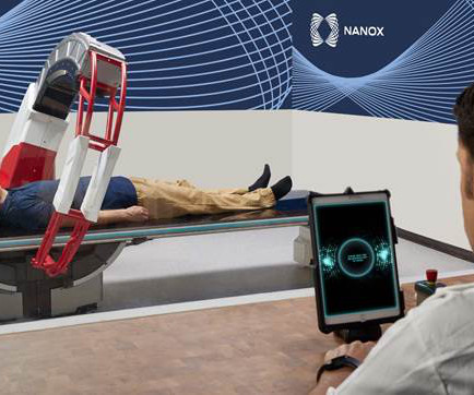
Approximately two-thirds of the world’s population does not have access to medical imaging systems, according to the World Health Organization ( WHO ),1 while many people with access face substantial wait times for scanning, potentially delaying diagnoses. While access to medical imaging is relatively high in the U.S.
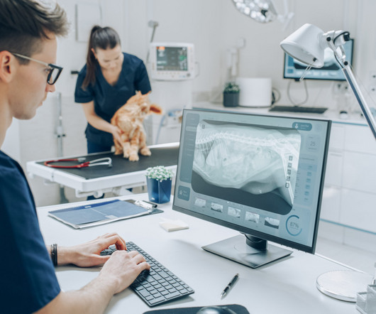
Veterinary technicians play a pivotal role in the radiographic process within veterinary practices. Their expertise in handling animals, positioning them correctly, and ensuring optimal image quality is essential for accurate diagnoses. Following strict safety protocols helps minimize risks.
We are excited about exploring the utility of pulmonary strain radiography towards diagnosing dyspnea.” Unlike with PFTs, our software enables us to generate pulmonary strain plots, comparable to strain echocardiography. Chest radiography is typically acquired in the evaluation of pulmonary disorders.
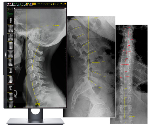
Lumbosacral spine X-rays, also called lumbar spine X-rays, are a radiographic imaging technique that uses low doses of electromagnetic radiation to view the internal anatomy of the lower spine, called the lumbosacral region. These images are used to diagnose a wide range of abnormalities, injuries, and diseases in the region.
Knowing that the industry continues to contend with staffing shortages and reduced budgets, Carestream is highlighting our advanced smart features that help radiographers execute quality exams in less time with less fatigue, leaving more time for patient interaction.
To date, 19 hospitals of Tripura have benefitted from Teleradiology services resulting in a total read-out of 61,660 radiographs by radiologists situated in Bangalore, Hyderabad, New Delhi, and other cities. The maximum number of radiographs reported were Chest radiographs(41.7%), followed by extremity radiographs(32.1%) for trauma.
A) AP radiograph of Lisfranc Fracture Dislocation demonstrates the circled “fleck sign” or Lisfranc ligament avulsion fracture fragment. (B) C) The lateral radiograph notes with a circle, the dorsal sub dislocation of the metatarsal base. of all diagnosed fractures. Trauma due to falling off a roof. Xray of the Week Figure 1.
A weight bearing CT scan can: Provide increased sensitivity and specificity over radiographs. Utility of WBCT to Diagnose Syndesmotic Instability in Patients With Weber B Lateral Malleolar Fractures. Help detect subtle syndesmosis injuries. PMID: 31442094. (2) MD; Waryasz, Gregory MD; DiGiovanni, Christopher W.
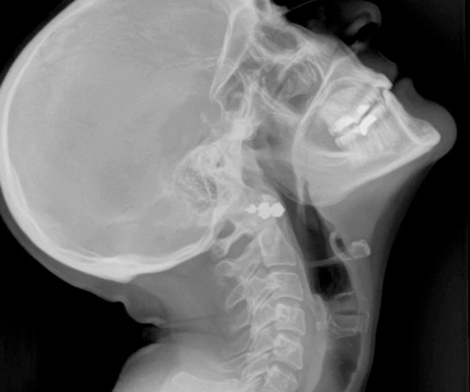
A patient’s specific needs and concerns are assessed, and a personalized radiographic plan is developed, taking into account factors like age, health conditions, and pregnancy. These methods provide comprehensive views of the oral cavity, enabling precise diagnoses and treatment planning.
The qXR-CTR is a deep-learning-based computer vision algorithm designed for use by physicians in all inpatient and outpatient settings, to automate the CTR assessment on chest radiographs (CXR). Prashant Warier , Co-Founder and CEO at Qure.ai, said, "We are delighted to receive FDA clearance for qXR-CTR.
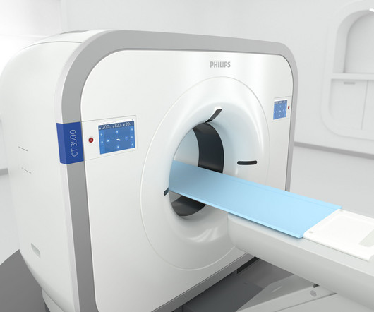
It automates radiographers’ most time-consuming steps so that they can spend more time focusing on the patient.” We’ve engineered the Philips CT 3500 to reduce the pain points that these high-volume departments face by developing a versatile, reliable, high-throughput imaging solution.
A radiographer is a medical professional who performs the scanning on patients. Nuclear medicine is used to diagnose some cancers, gastrointestinal issues, and endocrine disorders. Our fellowship-trained radiologists and radiology technicians are skilled at getting the results your doctor needs so you can be diagnosed accurately.
We organize all of the trending information in your field so you don't have to. Join 5,000 users and stay up to date on the latest articles your peers are reading.
You know about us, now we want to get to know you!
Let's personalize your content
Let's get even more personalized
We recognize your account from another site in our network, please click 'Send Email' below to continue with verifying your account and setting a password.


Let's personalize your content