This site uses cookies to improve your experience. To help us insure we adhere to various privacy regulations, please select your country/region of residence. If you do not select a country, we will assume you are from the United States. Select your Cookie Settings or view our Privacy Policy and Terms of Use.
Cookie Settings
Cookies and similar technologies are used on this website for proper function of the website, for tracking performance analytics and for marketing purposes. We and some of our third-party providers may use cookie data for various purposes. Please review the cookie settings below and choose your preference.
Used for the proper function of the website
Used for monitoring website traffic and interactions
Cookie Settings
Cookies and similar technologies are used on this website for proper function of the website, for tracking performance analytics and for marketing purposes. We and some of our third-party providers may use cookie data for various purposes. Please review the cookie settings below and choose your preference.
Strictly Necessary: Used for the proper function of the website
Performance/Analytics: Used for monitoring website traffic and interactions
12, 2025 Konica Minolta Healthcare Americas, has published a case study by clinicians in the pulmonary and radiology departments at ASST Fatebenefratelli Sacco (Milan, Italy) demonstrating the use of Dynamic Digital Radiography (DDR) to help definitively diagnose diaphragm dysfunction. tim.hodson Fri, 02/14/2025 - 15:14 Feb.12,
ChatGPT-4 outperformed human clinicians in determining pretest and post-test disease probability after a negative test result involving chest radiographs and mammograms, according to a research letter published December 11 in JAMA Network Open.
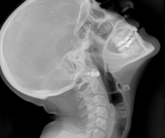
Chest dynamic digital radiography (DDR) may have received a boost toward clinical use in patients with lung disorders, with researchers developing AI to perform time-consuming analysis involved in the technology, according to researchers in New York City. a) Raw example of a dynamic digital radiograph. (b)
Researchers at the Icahn School of Medicine at Mount Sinai ("Icahn Mount Sinai") used Dynamic Digital Radiography (DDR) data, an X-ray imaging technology developed by Konica Minolta, to create their AI-powered technique that analyzes lung function. Chest radiography is typically acquired in the evaluation of pulmonary disorders.
It uses AI to analyze CT scans, X-rays and pathology slides, supporting clinicians in detecting and diagnosing medical conditions faster and more accurately. Averaged across all findings on chest radiographs.) [2]AIDE aims to address the global shortage of 1.5 Lancet Digital Health. Published 2021. 2]AIDE study, Alfred Health.
Morphological markers on MRI help diagnose autism. Commercially Available Chest Radiograph AI Tools for Detecting Airspace Disease, Pneumothorax, and Pleural Effusion. Generative Artificial Intelligence for Chest Radiograph Interpretation in the Emergency Department. Shah, et al, Radiography , January 9, 2024.
to demonstrate portable digital radiography and its effectiveness at high altitudes. Cairnie says, “Radiograph technology is critical for diagnosing several conditions, including bone fracture and chest conditions such as Tuberculosis. We were glad the expedition went well and now this technology is proven at high elevations."
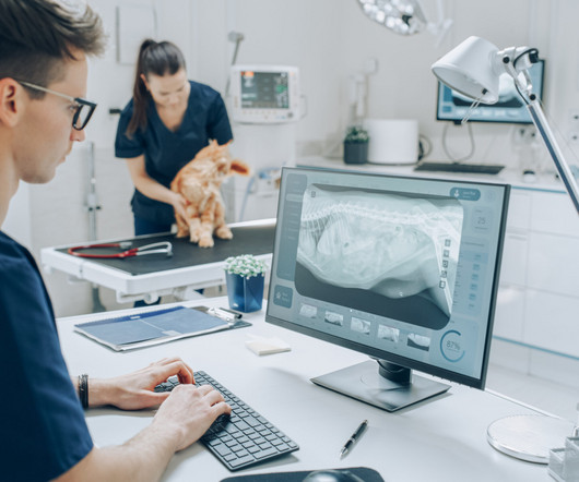
Veterinary technicians play a pivotal role in the radiographic process within veterinary practices. Their expertise in handling animals, positioning them correctly, and ensuring optimal image quality is essential for accurate diagnoses. Following strict safety protocols helps minimize risks.
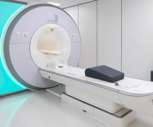
MRI-Scan-Teleradiology Introduction: Dental radiography is an essential component of modern dentistry, offering valuable insights into oral health and guiding treatments with precision. Patient-Centered Approach: Dental radiography starts with the patient. Quality Assurance: Dental radiographers are meticulous in quality assurance.
Key Points: Currently plain radiographs are the standard method in diagnosing syndesmotic ankle injuries even though the distal tibiofibular joint cannot be assessed due to superposition of the osseous structures in the foot.
Teleradiology Introduction: The landscape of chest radiography is undergoing a revolutionary transformation, with deep learning at its forefront. This blog post explores how deep learning algorithms are reshaping the future of chest radiography, particularly in the realm of pneumonia detection and classification.
The demands on radiography are growing in number and complexity, and Carestream is dedicated to delivering solutions that help solve our customers’ challenges,” said Vincent Chan , president and general manager of digital radiography at Carestream.
In the third blog of her series on AI and the radiographer, Shamie Kumar explores the impact on the radiographer when AI is integrated within an imaging modality. The question to explore in this blog is when AI is integrated within an imaging modality itself and how that may impact a radiographer.
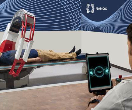
Nanox.ARC is a stationary X-ray system intended to produce tomographic images of the human musculoskeletal system adjunctive to conventional radiography on adult patients. Medical imaging is essential for detecting, diagnosing, and managing disease, guiding treatment decisions for improved health outcomes.
Again, while radiographers and radiologists are capturing and diagnosing images, the people signing off on the actual financial investment are the administrators, the CEOs, clinical engineering leaders, product committee chairs, and chief medical officers. Who is Making the Purchases?
Introduction: Dental X-ray technologists, also known as dental radiographers, are the unsung heroes behind the scenes of every successful dental diagnosis and treatment. Patient Interaction: Patient Comfort: Dental radiographers are responsible for ensuring the patient’s comfort and alleviating any anxiety.
A) AP radiograph of Lisfranc Fracture Dislocation demonstrates the circled “fleck sign” or Lisfranc ligament avulsion fracture fragment. (B) C) The lateral radiograph notes with a circle, the dorsal sub dislocation of the metatarsal base. of all diagnosed fractures. Trauma due to falling off a roof. Xray of the Week Figure 1.
Lens subluxation can be diagnosed by ultrasound which shows deviation of the lens (Fig. Radiography has no role in orbital injuries due to its lower sensitivity for soft tissues [4]. Radiographics. Trauma-Induced Bilateral Ectopia Lentis Diagnosed with Point-of-Care Ultrasound. 2006;10(4):345-350. doi: 10.1016/j.jaapos.2006.01.218
A growing number of medical organisations link to the DenseBreast-info.org website, including the EFRS (European Federation of Radiographer Societies) and the Society of Radiographers. Figure 4 (b) The website includes breast screening guidelines in Europe. A comparative analysis table summarises the guidelines in each country.
Trauma : In these injuries one would normally go for a standard radiography followed by second-line imaging such as CT. The possibility of inaccurate or missed diagnoses ranging on average from 20 to 40% in the literature is impossible to ignore and warrants 3D imaging as a primary modality.
Angela Young explains how the process of making a podcast helped not only others with a diagnosed brain tumour but gave comfort and support to herself as she embarked on a course of radiotherapy. and to many people from the team at Addenbrooke’s, including a medical physicist and a research radiographer.
We organize all of the trending information in your field so you don't have to. Join 5,000 users and stay up to date on the latest articles your peers are reading.
You know about us, now we want to get to know you!
Let's personalize your content
Let's get even more personalized
We recognize your account from another site in our network, please click 'Send Email' below to continue with verifying your account and setting a password.


Let's personalize your content